- PDB-1r5l: Crystal Structure of Human Alpha-Tocopherol Transfer Protein Boun... -
+
Open data
ID or keywords:
Loading...
-
Basic information
Entry
Database: PDB / ID: 1r5l
Title
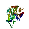
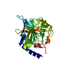
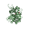
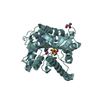
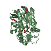
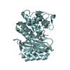
Crystal Structure of Human Alpha-Tocopherol Transfer Protein Bound to its Ligand
Components
PROTEIN (Alpha-tocopherol transfer protein)
Keywords
TRANSPORT PROTEIN / ATTP / TOCOPHEROL / ATAXIA WITH VITAMIN E DEFICIENCY
Function / homology
Function and homology information
Vitamin E transport / vitamin E binding / vitamin E metabolic process / vitamin transport / lipid transfer activity / intermembrane lipid transfer / negative regulation of establishment of blood-brain barrier / positive regulation of amyloid-beta clearance / phosphatidylinositol-3,4-bisphosphate binding / phosphatidylinositol bisphosphate binding ...Vitamin E transport / vitamin E binding / vitamin E metabolic process / vitamin transport / lipid transfer activity / intermembrane lipid transfer / negative regulation of establishment of blood-brain barrier / positive regulation of amyloid-beta clearance / phosphatidylinositol-3,4-bisphosphate binding / phosphatidylinositol bisphosphate binding / embryonic placenta development / phosphatidylinositol-4,5-bisphosphate binding / lipid metabolic process / response to toxic substance / late endosome / cytosol Similarity search - Function
Alpha-tocopherol transfer / N-terminal domain of phosphatidylinositol transfer protein sec14p / Phosphatidylinositol Transfer Protein Sec14p / CRAL-TRIO lipid binding domain / CRAL/TRIO, N-terminal domain / CRAL/TRIO, N-terminal domain / CRAL/TRIO, N-terminal domain / CRAL/TRIO, N-terminal domain superfamily / CRAL/TRIO domain / CRAL-TRIO lipid binding domain profile. ...Alpha-tocopherol transfer / N-terminal domain of phosphatidylinositol transfer protein sec14p / Phosphatidylinositol Transfer Protein Sec14p / CRAL-TRIO lipid binding domain / CRAL/TRIO, N-terminal domain / CRAL/TRIO, N-terminal domain / CRAL/TRIO, N-terminal domain / CRAL/TRIO, N-terminal domain superfamily / CRAL/TRIO domain / CRAL-TRIO lipid binding domain profile. / Domain in homologues of a S. cerevisiae phosphatidylinositol transfer protein (Sec14p) / CRAL-TRIO lipid binding domain / CRAL-TRIO lipid binding domain superfamily / Helicase, Ruva Protein; domain 3 / Single alpha-helices involved in coiled-coils or other helix-helix interfaces / Up-down Bundle / Orthogonal Bundle / 3-Layer(aba) Sandwich / Mainly Alpha / Alpha Beta Similarity search - Domain/homology
In the structure databanks used in Yorodumi, some data are registered as the other names, "COVID-19 virus" and "2019-nCoV". Here are the details of the virus and the list of structure data.
Jan 31, 2019. EMDB accession codes are about to change! (news from PDBe EMDB page)
EMDB accession codes are about to change! (news from PDBe EMDB page)
The allocation of 4 digits for EMDB accession codes will soon come to an end. Whilst these codes will remain in use, new EMDB accession codes will include an additional digit and will expand incrementally as the available range of codes is exhausted. The current 4-digit format prefixed with “EMD-” (i.e. EMD-XXXX) will advance to a 5-digit format (i.e. EMD-XXXXX), and so on. It is currently estimated that the 4-digit codes will be depleted around Spring 2019, at which point the 5-digit format will come into force.
The EM Navigator/Yorodumi systems omit the EMD- prefix.
Related info.:Q: What is EMD? / ID/Accession-code notation in Yorodumi/EM Navigator
Yorodumi is a browser for structure data from EMDB, PDB, SASBDB, etc.
This page is also the successor to EM Navigator detail page, and also detail information page/front-end page for Omokage search.
The word "yorodu" (or yorozu) is an old Japanese word meaning "ten thousand". "mi" (miru) is to see.
Related info.:EMDB / PDB / SASBDB / Comparison of 3 databanks / Yorodumi Search / Aug 31, 2016. New EM Navigator & Yorodumi / Yorodumi Papers / Jmol/JSmol / Function and homology information / Changes in new EM Navigator and Yorodumi
 Movie
Movie Controller

 Yorodumi
 Open data
 Basic information
 Components
 Keywords
 Function and homology information
 Homo sapiens (human)
 X-RAY DIFFRACTION /
  Authors
 Citation
 Structure visualization
 Downloads & links
 Other downloads
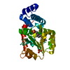


 PDBj
PDBj Assembly
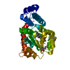

 Mass: 430.706 Da / Num. of mol.: 1 / Source method: obtained synthetically / Formula: C29H50O2
 Mass: 18.015 Da / Num. of mol.: 253 / Source method: isolated from a natural source / Formula: H2O
 Sample preparation
 Processing