[English] 日本語
Yorodumi
- PDB-3vc1: Crystal structure of geranyl diphosphate C-methyltransferase from... -

+
Open data


ID or keywords:

Loading...

-
Basic information

Entry
Database: PDB / ID: 3vc1
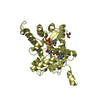
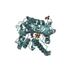
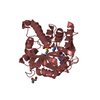
Title
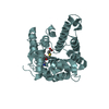
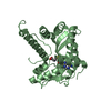
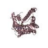
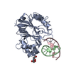
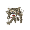
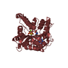
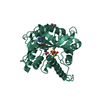
Crystal structure of geranyl diphosphate C-methyltransferase from Streptomyces coelicolor A3(2) in complex with Mg2+, geranyl-S-thiolodiphosphate, and S-adenosyl-L-homocysteine
ComponentsGeranyl diphosphate 2-C-methyltransferase
KeywordsTRANSFERASE / Rossmann fold / methyltransferase fold / SAM-dependent methyltransferase / C-methyltransferase / terpenoid biosynthesis / 2-methylisoborneol biosynthesis
Function / homology
Function and homology information


geranyl diphosphate 2-C-methyltransferase / C-methyltransferase activity / terpene metabolic process / S-adenosyl-L-methionine binding / S-adenosylmethionine-dependent methyltransferase activity / methylation / magnesium ion binding
Similarity search - Function
: / Methyltransferase type 11 / Methyltransferase domain / Vaccinia Virus protein VP39 / S-adenosyl-L-methionine-dependent methyltransferase superfamily / Rossmann fold / 3-Layer(aba) Sandwich / Alpha Beta
Similarity search - Domain/homology
GERANYL S-THIOLODIPHOSPHATE / S-ADENOSYL-L-HOMOCYSTEINE / Geranyl diphosphate 2-C-methyltransferase
Similarity search - Component
Biological speciesStreptomyces coelicolor (bacteria)
MethodX-RAY DIFFRACTION / SYNCHROTRON / SIRAS / Resolution: 1.82 Å
AuthorsKoksal, M. / Christianson, D.W.
CitationJournal: Biochemistry / Year: 2012
Title: Structure of Geranyl Diphosphate C-Methyltransferase from Streptomyces coelicolor and Implications for the Mechanism of Isoprenoid Modification.
Authors: Koksal, M. / Chou, W.K. / Cane, D.E. / Christianson, D.W.
History
DepositionJan 3, 2012Deposition site: RCSB / Processing site: RCSB
Revision 1.0Apr 11, 2012Provider: repository / Type: Initial release
Revision 1.1Apr 25, 2012Group: Database references
Revision 1.2Nov 8, 2017Group: Refinement description / Category: software / Item: _software.name
Revision 1.3Feb 28, 2024Group: Data collection / Database references / Derived calculations
Category: chem_comp_atom / chem_comp_bond ...chem_comp_atom / chem_comp_bond / database_2 / pdbx_struct_conn_angle / struct_conn / struct_ref_seq_dif / struct_site
Item: _database_2.pdbx_DOI / _database_2.pdbx_database_accession ..._database_2.pdbx_DOI / _database_2.pdbx_database_accession / _pdbx_struct_conn_angle.ptnr1_auth_asym_id / _pdbx_struct_conn_angle.ptnr1_auth_comp_id / _pdbx_struct_conn_angle.ptnr1_auth_seq_id / _pdbx_struct_conn_angle.ptnr1_label_asym_id / _pdbx_struct_conn_angle.ptnr1_label_atom_id / _pdbx_struct_conn_angle.ptnr1_label_comp_id / _pdbx_struct_conn_angle.ptnr1_label_seq_id / _pdbx_struct_conn_angle.ptnr2_auth_asym_id / _pdbx_struct_conn_angle.ptnr2_label_asym_id / _pdbx_struct_conn_angle.ptnr3_auth_asym_id / _pdbx_struct_conn_angle.ptnr3_auth_comp_id / _pdbx_struct_conn_angle.ptnr3_auth_seq_id / _pdbx_struct_conn_angle.ptnr3_label_asym_id / _pdbx_struct_conn_angle.ptnr3_label_atom_id / _pdbx_struct_conn_angle.ptnr3_label_comp_id / _pdbx_struct_conn_angle.ptnr3_label_seq_id / _pdbx_struct_conn_angle.value / _struct_conn.pdbx_dist_value / _struct_conn.ptnr1_auth_asym_id / _struct_conn.ptnr1_auth_comp_id / _struct_conn.ptnr1_auth_seq_id / _struct_conn.ptnr1_label_asym_id / _struct_conn.ptnr1_label_atom_id / _struct_conn.ptnr1_label_comp_id / _struct_conn.ptnr1_label_seq_id / _struct_conn.ptnr2_auth_asym_id / _struct_conn.ptnr2_auth_comp_id / _struct_conn.ptnr2_auth_seq_id / _struct_conn.ptnr2_label_asym_id / _struct_conn.ptnr2_label_atom_id / _struct_conn.ptnr2_label_comp_id / _struct_ref_seq_dif.details / _struct_site.pdbx_auth_asym_id / _struct_site.pdbx_auth_comp_id / _struct_site.pdbx_auth_seq_id

-
Structure visualization

Structure viewerMolecule:
MolmilJmol/JSmol

Downloads & links

-
Assembly

Deposited unit
A: Geranyl diphosphate 2-C-methyltransferase
B: Geranyl diphosphate 2-C-methyltransferase
C: Geranyl diphosphate 2-C-methyltransferase
D: Geranyl diphosphate 2-C-methyltransferase
E: Geranyl diphosphate 2-C-methyltransferase
F: Geranyl diphosphate 2-C-methyltransferase
G: Geranyl diphosphate 2-C-methyltransferase
H: Geranyl diphosphate 2-C-methyltransferase
I: Geranyl diphosphate 2-C-methyltransferase
J: Geranyl diphosphate 2-C-methyltransferase
K: Geranyl diphosphate 2-C-methyltransferase
L: Geranyl diphosphate 2-C-methyltransferase
hetero molecules


Theoretical massNumber of molelcules
Total (without water)430,82664
Polymers420,48112
Non-polymers10,34552
Water34,3011904
1
A: Geranyl diphosphate 2-C-methyltransferase
hetero molecules


Theoretical massNumber of molelcules
Total (without water)36,0557
Polymers35,0401
Non-polymers1,0156
Water181
TypeNameSymmetry operationNumber
identity operation1_555x,y,z1
MethodPISA
2
B: Geranyl diphosphate 2-C-methyltransferase
hetero molecules


Theoretical massNumber of molelcules
Total (without water)35,8715
Polymers35,0401
Non-polymers8314
Water181
TypeNameSymmetry operationNumber
identity operation1_555x,y,z1
MethodPISA
3
C: Geranyl diphosphate 2-C-methyltransferase
hetero molecules


Theoretical massNumber of molelcules
Total (without water)35,9636
Polymers35,0401
Non-polymers9235
Water181
TypeNameSymmetry operationNumber
identity operation1_555x,y,z1
MethodPISA
4
D: Geranyl diphosphate 2-C-methyltransferase
hetero molecules


Theoretical massNumber of molelcules
Total (without water)35,8715
Polymers35,0401
Non-polymers8314
Water181
TypeNameSymmetry operationNumber
identity operation1_555x,y,z1
MethodPISA
5
E: Geranyl diphosphate 2-C-methyltransferase
hetero molecules


Theoretical massNumber of molelcules
Total (without water)36,0597
Polymers35,0401
Non-polymers1,0196
Water181
TypeNameSymmetry operationNumber
identity operation1_555x,y,z1
MethodPISA
6
F: Geranyl diphosphate 2-C-methyltransferase
hetero molecules


Theoretical massNumber of molelcules
Total (without water)35,8715
Polymers35,0401
Non-polymers8314
Water181
TypeNameSymmetry operationNumber
identity operation1_555x,y,z1
MethodPISA
7
G: Geranyl diphosphate 2-C-methyltransferase
hetero molecules


Theoretical massNumber of molelcules
Total (without water)35,8715
Polymers35,0401
Non-polymers8314
Water181
TypeNameSymmetry operationNumber
identity operation1_555x,y,z1
MethodPISA
8
H: Geranyl diphosphate 2-C-methyltransferase
hetero molecules


Theoretical massNumber of molelcules
Total (without water)35,8715
Polymers35,0401
Non-polymers8314
Water181
TypeNameSymmetry operationNumber
identity operation1_555x,y,z1
MethodPISA
9
I: Geranyl diphosphate 2-C-methyltransferase
hetero molecules


Theoretical massNumber of molelcules
Total (without water)35,8715
Polymers35,0401
Non-polymers8314
Water181
TypeNameSymmetry operationNumber
identity operation1_555x,y,z1
MethodPISA
10
J: Geranyl diphosphate 2-C-methyltransferase
hetero molecules


Theoretical massNumber of molelcules
Total (without water)35,8715
Polymers35,0401
Non-polymers8314
Water181
TypeNameSymmetry operationNumber
identity operation1_555x,y,z1
MethodPISA
11
K: Geranyl diphosphate 2-C-methyltransferase
hetero molecules


Theoretical massNumber of molelcules
Total (without water)35,7794
Polymers35,0401
Non-polymers7393
Water181
TypeNameSymmetry operationNumber
identity operation1_555x,y,z1
MethodPISA
12
L: Geranyl diphosphate 2-C-methyltransferase
hetero molecules


Theoretical massNumber of molelcules
Total (without water)35,8715
Polymers35,0401
Non-polymers8314
Water181
TypeNameSymmetry operationNumber
identity operation1_555x,y,z1
MethodPISA
Unit cell
Length a, b, c (Å)97.029, 102.903, 203.582
Angle α, β, γ (deg.)90.00, 99.59, 90.00
Int Tables number4
Space group name H-MP1211

-
Components

-
Protein , 1 types, 12 molecules ABCDEFGHIJKL

#1: Protein
Geranyl diphosphate 2-C-methyltransferase / / GPP methyltransferase


Mass: 35040.090 Da / Num. of mol.: 12
Source method: isolated from a genetically manipulated source
Source: (gene. exp.) Streptomyces coelicolor (bacteria) / Strain: A3(2) / Gene: orf4, SCBAC12C8.02, SCO7701 / Plasmid: pET28a / Production host: Escherichia coli (E. coli) / Strain (production host): BL21(DE3)
References: UniProt: Q9F1Y5, Transferases; Transferring one-carbon groups; Methyltransferases

-
Non-polymers , 6 types, 1956 molecules

#2: Chemical
ChemComp-MG / MAGNESIUM ION


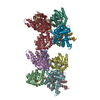
Mass: 24.305 Da / Num. of mol.: 12 / Source method: obtained synthetically / Formula: Mg
#3: Chemical
ChemComp-SAH / S-ADENOSYL-L-HOMOCYSTEINE / S-Adenosyl-L-homocysteine


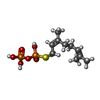
Type: L-peptide linking / Mass: 384.411 Da / Num. of mol.: 12 / Source method: obtained synthetically / Formula: C14H20N6O5S
#4: Chemical
ChemComp-GST / GERANYL S-THIOLODIPHOSPHATE / S-[(2E)-3,7-DIMETHYLOCTA-2,6-DIENYL] TRIHYDROGEN THIODIPHOSPHATE


Mass: 330.275 Da / Num. of mol.: 12 / Source method: obtained synthetically / Formula: C10H20O6P2S
#5: Chemical
ChemComp-GOL / GLYCEROL / GLYCERIN / PROPANE-1,2,3-TRIOL / Glycerol


Mass: 92.094 Da / Num. of mol.: 15 / Source method: obtained synthetically / Formula: C3H8O3
#6: Chemical ChemComp-SO4 / SULFATE ION / Sulfate


Mass: 96.063 Da / Num. of mol.: 1 / Source method: obtained synthetically / Formula: SO4
#7: Water ChemComp-HOH / water / Water


Mass: 18.015 Da / Num. of mol.: 1904 / Source method: isolated from a natural source / Formula: H2O

-
Experimental details

-
Experiment

ExperimentMethod: X-RAY DIFFRACTION / Number of used crystals: 1

-
Sample preparation

CrystalDensity Matthews: 2.38 Å3/Da / Density % sol: 48.39 %
Crystal growTemperature: 294 K / Method: vapor diffusion, sitting drop / pH: 7.5
Details: 100 mM HEPES (pH 7.5), 25% polyethylene glycol 3350, 200 mM (NH4)2SO4, VAPOR DIFFUSION, SITTING DROP, temperature 294K

-
Data collection

DiffractionMean temperature: 100 K
Diffraction sourceSource: SYNCHROTRON / Site: NSLS / Beamline: X29A / Wavelength: 1.075 Å
DetectorType: ADSC QUANTUM 315r / Detector: CCD / Date: Nov 3, 2011
Details: Cryogenically cooled double crystal monochrometer with horizontal focusing sagittal bend second mono crystal with 4:1 magnification ratio and vertically focusing mirror
RadiationMonochromator: Double silicon(111) crystal / Protocol: SINGLE WAVELENGTH / Monochromatic (M) / Laue (L): M / Scattering type: x-ray
Radiation wavelengthWavelength: 1.075 Å / Relative weight: 1
ReflectionResolution: 1.82→50 Å / Num. all: 372879 / Num. obs: 348804 / % possible obs: 99.1 % / Observed criterion σ(F): 0 / Observed criterion σ(I): -3 / Redundancy: 3.1 % / Rmerge(I) obs: 0.106 / Rsym value: 0.106 / Net I/σ(I): 10.2
Reflection shellResolution: 1.82→1.89 Å / Redundancy: 2.9 % / Rmerge(I) obs: 0.536 / Mean I/σ(I) obs: 2.1 / Num. unique all: 34608 / Rsym value: 0.536 / % possible all: 98.7

-
Processing

Software
NameVersionClassification
CBASSdata collection
HKL2Mapmodel building
PHENIX(phenix.refine: 1.5_2)refinement
HKL-2000data reduction
HKL-2000data scaling
HKL2Mapphasing
RefinementMethod to determine structure: SIRAS / Resolution: 1.82→46.28 Å / SU ML: 0.18 / σ(F): 0.07 / Phase error: 19.46 / Stereochemistry target values: ML
RfactorNum. reflection% reflectionSelection details
Rfree0.1936 1859 57 %RANDOM
Rwork0.1667 ---
all0.1669 348804 --
obs0.1669 324579 92.04 %-
Solvent computationShrinkage radii: 0.9 Å / VDW probe radii: 1.11 Å / Solvent model: FLAT BULK SOLVENT MODEL / Bsol: 40.024 Å2 / ksol: 0.4 e/Å3
Displacement parameters
Baniso -1Baniso -2Baniso -3
1-5.8366 Å2-0 Å22.7913 Å2
2---2.2001 Å20 Å2
3----3.6365 Å2
Refine analyze
FreeObs
Luzzati coordinate error0.19 Å0.19 Å
Luzzati d res low-5 Å
Luzzati sigma a0.18 Å0.18 Å
Refinement stepCycle: LAST / Resolution: 1.82→46.28 Å
ProteinNucleic acidLigandSolventTotal
Num. atoms26616 0 647 1904 29167
Refine LS restraints
Refine-IDTypeDev idealNumber
X-RAY DIFFRACTIONf_bond_d0.01627965
X-RAY DIFFRACTIONf_angle_d1.61137961
X-RAY DIFFRACTIONf_dihedral_angle_d17.92610125
X-RAY DIFFRACTIONf_chiral_restr0.113960
X-RAY DIFFRACTIONf_plane_restr0.0084979
LS refinement shell
Resolution (Å)Rfactor RfreeNum. reflection RfreeRfactor RworkNum. reflection RworkRefine-ID% reflection obs (%)
1.8203-1.86950.26211220.234721095X-RAY DIFFRACTION78
1.8695-1.92450.23691320.217322890X-RAY DIFFRACTION85
1.9245-1.98660.27391360.202723681X-RAY DIFFRACTION88
1.9866-2.05760.22791380.183124225X-RAY DIFFRACTION90
2.0576-2.140.22281510.172824606X-RAY DIFFRACTION92
2.14-2.23740.21341390.170924942X-RAY DIFFRACTION93
2.2374-2.35540.19811440.166525176X-RAY DIFFRACTION94
2.3554-2.50290.20791490.171525432X-RAY DIFFRACTION95
2.5029-2.69620.19961470.165525841X-RAY DIFFRACTION96
2.6962-2.96740.20111470.171525998X-RAY DIFFRACTION96
2.9674-3.39670.17761530.168126019X-RAY DIFFRACTION96
3.3967-4.2790.15911500.141525675X-RAY DIFFRACTION95
4.279-46.29520.16611510.149627140X-RAY DIFFRACTION99

+
About Yorodumi

-
News

-
Feb 9, 2022. New format data for meta-information of EMDB entries

New format data for meta-information of EMDB entries

  • Version 3 of the EMDB header file is now the official format.
  • The previous official version 1.9 will be removed from the archive.

Related info.:EMDB header

External links:wwPDB to switch to version 3 of the EMDB data model

-
Aug 12, 2020. Covid-19 info

Covid-19 info

URL: https://pdbj.org/emnavi/covid19.php

New page: Covid-19 featured information page in EM Navigator.

Related info.:Covid-19 info / Mar 5, 2020. Novel coronavirus structure data

+
Mar 5, 2020. Novel coronavirus structure data

Novel coronavirus structure data

Related info.:Yorodumi Speices / Aug 12, 2020. Covid-19 info

External links:COVID-19 featured content - PDBj / Molecule of the Month (242):Coronavirus Proteases

+
Jan 31, 2019. EMDB accession codes are about to change! (news from PDBe EMDB page)

EMDB accession codes are about to change! (news from PDBe EMDB page)

  • The allocation of 4 digits for EMDB accession codes will soon come to an end. Whilst these codes will remain in use, new EMDB accession codes will include an additional digit and will expand incrementally as the available range of codes is exhausted. The current 4-digit format prefixed with “EMD-” (i.e. EMD-XXXX) will advance to a 5-digit format (i.e. EMD-XXXXX), and so on. It is currently estimated that the 4-digit codes will be depleted around Spring 2019, at which point the 5-digit format will come into force.
  • The EM Navigator/Yorodumi systems omit the EMD- prefix.

Related info.:Q: What is EMD? / ID/Accession-code notation in Yorodumi/EM Navigator

External links:EMDB Accession Codes are Changing Soon! / Contact to PDBj

+
Jul 12, 2017. Major update of PDB

Major update of PDB

  • wwPDB released updated PDB data conforming to the new PDBx/mmCIF dictionary.
  • This is a major update changing the version number from 4 to 5, and with Remediation, in which all the entries are updated.
  • In this update, many items about electron microscopy experimental information are reorganized (e.g. em_software).
  • Now, EM Navigator and Yorodumi are based on the updated data.

External links:wwPDB Remediation / Enriched Model Files Conforming to OneDep Data Standards Now Available in the PDB FTP Archive

-
Yorodumi

Thousand views of thousand structures

  • Yorodumi is a browser for structure data from EMDB, PDB, SASBDB, etc.
  • This page is also the successor to EM Navigator detail page, and also detail information page/front-end page for Omokage search.
  • The word "yorodu" (or yorozu) is an old Japanese word meaning "ten thousand". "mi" (miru) is to see.

Related info.:EMDB / PDB / SASBDB / Comparison of 3 databanks / Yorodumi Search / Aug 31, 2016. New EM Navigator & Yorodumi / Yorodumi Papers / Jmol/JSmol / Function and homology information / Changes in new EM Navigator and Yorodumi

Read more