 Movie
Movie Controller

[English] 日本語
 Yorodumi
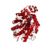
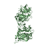
- PDB-2dnj: DNASE I-INDUCED DNA CONFORMATION. 2 ANGSTROMS STRUCTURE OF A DNAS... -
+ Open data

- Basic information
| Entry | Database: PDB / ID: 2dnj | |||||||||
|---|---|---|---|---|---|---|---|---|---|---|
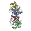
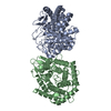
| Title | DNASE I-INDUCED DNA CONFORMATION. 2 ANGSTROMS STRUCTURE OF A DNASE I-OCTAMER COMPLEX | |||||||||
 Components Components |
| |||||||||
 Keywords Keywords | HYDROLASE/DNA / PROTEIN-DNA COMPLEX / DOUBLE HELIX / HYDROLASE-DNA COMPLEX | |||||||||
| Function / homology |  Function and homology information Function and homology informationregulation of neutrophil mediated cytotoxicity / zymogen granule / regulation of acute inflammatory response / deoxyribonuclease I / neutrophil activation involved in immune response / deoxyribonuclease I activity / DNA catabolic process / nuclear envelope / actin binding / apoptotic process ...regulation of neutrophil mediated cytotoxicity / zymogen granule / regulation of acute inflammatory response / deoxyribonuclease I / neutrophil activation involved in immune response / deoxyribonuclease I activity / DNA catabolic process / nuclear envelope / actin binding / apoptotic process / DNA binding / extracellular region / nucleus Similarity search - Function | |||||||||
| Biological species |  | |||||||||
| Method |  X-RAY DIFFRACTION / Resolution: 2 Å X-RAY DIFFRACTION / Resolution: 2 Å | |||||||||
 Authors Authors | Lahm, A. / Suck, D. | |||||||||
 Citation Citation | Journal: J.Mol.Biol. / Year: 1991 Title: DNase I-induced DNA conformation. 2 A structure of a DNase I-octamer complex Authors: Lahm, A. / Suck, D. #1: Journal: Nature / Year: 1988Title: Structure Refined to 2 Angstroms of a Nicked DNA Octanucleotide Complex with DNase I Authors: Suck, D. / Lahm, A. / Oefner, C. #2: Journal: Nature / Year: 1986Title: Structure of DNase I at 2.0 Angstroms Resolution Suggests a Mechanism for Binding to and Cutting DNA Authors: Suck, D. / Oefner, C. #3: Journal: Embo J. / Year: 1984Title: Three-Dimensional Structure of Bovine Pancreatic DNase I at 2.5 Angstroms Resolution Authors: Suck, D. / Oefner, C. / Kabsch, W. #4: Journal: J.Mol.Biol. / Year: 1982Title: Crystallization and Preliminary Crystallographic Data of Bovine Pancreatic Deoxyribonuclease I Authors: Suck, D. #5: Journal: J.Biol.Chem. / Year: 1973Title: Bovine Pancreatic Deoxyribonuclease A. Isolation of Cyanogen Bromide Peptides, Complete Covalent Structure of the Polypeptide Chain Authors: Liao, T.-H. / Salnikow, J. / Moore, S. / Stein, W.H. | |||||||||
| History |
|
- Structure visualization
| Structure viewer | Molecule: MolmilJmol/JSmol |
|---|
- Downloads & links
-Download
| PDBx/mmCIF format | 2dnj.cif.gz | 81 KB | Display | PDBx/mmCIF format |
|---|---|---|---|---|
| PDB format | pdb2dnj.ent.gz | 56.9 KB | Display | PDB format |
| PDBx/mmJSON format | 2dnj.json.gz | Tree view | PDBx/mmJSON format | |
| Others |  Other downloads Other downloads |
-Validation report
| Arichive directory | https://data.pdbj.org/pub/pdb/validation_reports/dn/2dnjftp://data.pdbj.org/pub/pdb/validation_reports/dn/2dnj | HTTPS FTP |
|---|
-Related structure data
| Similar structure data |
|---|


-Links
 PDBj
PDBj

- Assembly
| Deposited unit | 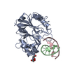
| ||||||||||
|---|---|---|---|---|---|---|---|---|---|---|---|
| 1 | 
| ||||||||||
| Unit cell |
| ||||||||||
| Details | THE ASYMMETRIC UNIT CONTAINS ONE DNASE I MOLECULE AND ONE NICKED DNA OCTAMER WITH A TOTAL OF 14 NUICLEOTIDES. THE CLEAVED 3-PRIME GC OF ONE OF THE DNA STRANDS IS MISSING IN THE STRUCTURE. THE SYMMETRY OPERATION X,1-Y,1-Z, I.E. THE TWOFOLD AXIS ALONG A AT B/2,C/2, CREATES THE QUASI-CONTINUOUS 14-MER FORMED BY THE TWO OVERLAPPING NICKE OCTAMERS. |
-Components
| #1: DNA chain | Mass: 2427.605 Da / Num. of mol.: 1 / Source method: obtained synthetically |
|---|---|
| #2: DNA chain | Mass: 1809.218 Da / Num. of mol.: 1 / Source method: obtained synthetically |
| #3: Protein | Mass: 29092.574 Da / Num. of mol.: 1 / Source method: isolated from a natural source / Source: (natural) |
| #4: Sugar | ChemComp-NAG /   Type: D-saccharide, beta linking / Mass: 221.208 Da / Num. of mol.: 1 Type: D-saccharide, beta linking / Mass: 221.208 Da / Num. of mol.: 1Source method: isolated from a genetically manipulated source Formula: C8H15NO6 |
| #5: Water | ChemComp-HOH /  Mass: 18.015 Da / Num. of mol.: 252 / Source method: isolated from a natural source / Formula: H2O Mass: 18.015 Da / Num. of mol.: 252 / Source method: isolated from a natural source / Formula: H2O |
| Has protein modification | Y |
| Nonpolymer details | OF THE CARBOHYDRATE MOIETY ATTACHED TO ND2 ASN 18, ONLY THE FIRST RESIDUE WAS VISIBLE IN THE ...OF THE CARBOHYDRA |
-Experimental details
-Experiment
| Experiment | Method: X-RAY DIFFRACTION |
|---|
- Sample preparation
| Crystal | Density Matthews: 2.52 Å3/Da / Density % sol: 51.17 % | ||||||||||||||||||||||||
|---|---|---|---|---|---|---|---|---|---|---|---|---|---|---|---|---|---|---|---|---|---|---|---|---|---|
| Crystal grow | Temperature: 277 K / Method: vapor diffusion / pH: 6 / Details: pH 6.00, VAPOR DIFFUSION, temperature 277.00K | ||||||||||||||||||||||||
| Components of the solutions |
| ||||||||||||||||||||||||
| Crystal grow | *PLUS Temperature: 4 ℃ / pH: 6 / Method: other | ||||||||||||||||||||||||
| Components of the solutions | *PLUS
|
-Data collection
| Diffraction | Mean temperature: 273 K |
|---|---|
| Diffraction source | Source: ROTATING ANODE |
| Detector | Detector: OSCILLATION CAMERA |
| Radiation | Protocol: SINGLE WAVELENGTH / Monochromatic (M) / Laue (L): M / Scattering type: x-ray |
| Radiation wavelength | Relative weight: 1 |
| Reflection | Resolution: 2→50 Å / Num. obs: 20921 / Observed criterion σ(F): 3 |
| Reflection | *PLUS Highest resolution: 2 Å / Lowest resolution: 50 Å / % possible obs: 89.2 % / Rmerge(I) obs: 0.076 |
- Processing
| Software | Name: PROLSQ / Classification: refinement | ||||||||||||||||||||||||||||||||||||||||||||||||||||||||||||||||||||||||||||||||||||
|---|---|---|---|---|---|---|---|---|---|---|---|---|---|---|---|---|---|---|---|---|---|---|---|---|---|---|---|---|---|---|---|---|---|---|---|---|---|---|---|---|---|---|---|---|---|---|---|---|---|---|---|---|---|---|---|---|---|---|---|---|---|---|---|---|---|---|---|---|---|---|---|---|---|---|---|---|---|---|---|---|---|---|---|---|---|
| Refinement | Resolution: 2→6 Å / σ(F): 0 /
| ||||||||||||||||||||||||||||||||||||||||||||||||||||||||||||||||||||||||||||||||||||
| Refinement step | Cycle: LAST / Resolution: 2→6 Å
| ||||||||||||||||||||||||||||||||||||||||||||||||||||||||||||||||||||||||||||||||||||
| Refine LS restraints |
| ||||||||||||||||||||||||||||||||||||||||||||||||||||||||||||||||||||||||||||||||||||
| Software | *PLUS Name: PROLSQ / Classification: refinement | ||||||||||||||||||||||||||||||||||||||||||||||||||||||||||||||||||||||||||||||||||||
| Refinement | *PLUS Highest resolution: 2 Å / Lowest resolution: 6 Å / Num. reflection all: 20101 / σ(F): 0 / Rfactor all: 0.174 | ||||||||||||||||||||||||||||||||||||||||||||||||||||||||||||||||||||||||||||||||||||
| Solvent computation | *PLUS | ||||||||||||||||||||||||||||||||||||||||||||||||||||||||||||||||||||||||||||||||||||
| Displacement parameters | *PLUS |
