 Movie
Movie Controller


+ Open data

- Basic information
| Entry | Database: PDB / ID: 1cjb | ||||||
|---|---|---|---|---|---|---|---|
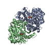
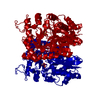
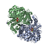
| Title | MALARIAL PURINE PHOSPHORIBOSYLTRANSFERASE | ||||||
 Components Components | PROTEIN (HYPOXANTHINE-GUANINE PHOSPHORIBOSYLTRANSFERASE) | ||||||
 Keywords Keywords | TRANSFERASE / MALARIA / PURINE SALVAGE / PHOSPHORIBOSYLTRANSFERASE / TRANSITION STATE INHIBITOR | ||||||
| Function / homology |  Function and homology information Function and homology informationxanthine phosphoribosyltransferase / XMP salvage / xanthine phosphoribosyltransferase activity / hypoxanthine phosphoribosyltransferase / guanine phosphoribosyltransferase activity / guanine salvage / hypoxanthine metabolic process / hypoxanthine phosphoribosyltransferase activity / GMP salvage / IMP salvage ...xanthine phosphoribosyltransferase / XMP salvage / xanthine phosphoribosyltransferase activity / hypoxanthine phosphoribosyltransferase / guanine phosphoribosyltransferase activity / guanine salvage / hypoxanthine metabolic process / hypoxanthine phosphoribosyltransferase activity / GMP salvage / IMP salvage / purine ribonucleoside salvage / nucleotide binding / magnesium ion binding / cytosol Similarity search - Function | ||||||
| Biological species |  | ||||||
| Method |  X-RAY DIFFRACTION / SYNCHROTRON / MOLECULAR REPLACEMENT / Resolution: 2 Å X-RAY DIFFRACTION / SYNCHROTRON / MOLECULAR REPLACEMENT / Resolution: 2 Å | ||||||
 Authors Authors | Shi, W. / Li, C.M. / Tyler, P.C. / Furneaux, R.H. / Cahill, S.M. / Girvin, M.E. / Grubmeyer, C. / Schramm, V.L. / Almo, S.C. | ||||||
 Citation Citation | Journal: Biochemistry / Year: 1999 Title: The 2.0 A structure of malarial purine phosphoribosyltransferase in complex with a transition-state analogue inhibitor. Authors: Shi, W. / Li, C.M. / Tyler, P.C. / Furneaux, R.H. / Cahill, S.M. / Girvin, M.E. / Grubmeyer, C. / Schramm, V.L. / Almo, S.C. | ||||||
| History |
|
- Structure visualization
| Structure viewer | Molecule: MolmilJmol/JSmol |
|---|
- Downloads & links
-Download
| PDBx/mmCIF format | 1cjb.cif.gz | 203.7 KB | Display | PDBx/mmCIF format |
|---|---|---|---|---|
| PDB format | pdb1cjb.ent.gz | 162.4 KB | Display | PDB format |
| PDBx/mmJSON format | 1cjb.json.gz | Tree view | PDBx/mmJSON format | |
| Others |  Other downloads Other downloads |
-Validation report
| Arichive directory | https://data.pdbj.org/pub/pdb/validation_reports/cj/1cjbftp://data.pdbj.org/pub/pdb/validation_reports/cj/1cjb | HTTPS FTP |
|---|
-Related structure data
| Related structure data |  1bzyS S: Starting model for refinement |
|---|---|
| Similar structure data |


-Links
 PDBj
PDBj


- Assembly
| Deposited unit | 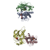
| ||||||||||||||||
|---|---|---|---|---|---|---|---|---|---|---|---|---|---|---|---|---|---|
| 1 | 
| ||||||||||||||||
| 2 | 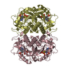
| ||||||||||||||||
| Unit cell |
| ||||||||||||||||
| Noncrystallographic symmetry (NCS) | NCS oper:
|
-Components
| #1: Protein | Mass: 26386.457 Da / Num. of mol.: 4 Source method: isolated from a genetically manipulated source Source: (gene. exp.) Production host:  References: UniProt: P20035, hypoxanthine phosphoribosyltransferase #2: Chemical | ChemComp-MG /   Mass: 24.305 Da / Num. of mol.: 8 / Source method: obtained synthetically / Formula: Mg Mass: 24.305 Da / Num. of mol.: 8 / Source method: obtained synthetically / Formula: Mg#3: Chemical | ChemComp-IRP / (   Mass: 346.233 Da / Num. of mol.: 4 / Source method: obtained synthetically / Formula: C11H15N4O7P Mass: 346.233 Da / Num. of mol.: 4 / Source method: obtained synthetically / Formula: C11H15N4O7P#4: Chemical | ChemComp-POP /   Mass: 175.959 Da / Num. of mol.: 4 / Source method: obtained synthetically / Formula: H2O7P2 Mass: 175.959 Da / Num. of mol.: 4 / Source method: obtained synthetically / Formula: H2O7P2#5: Water | ChemComp-HOH / |  Mass: 18.015 Da / Num. of mol.: 407 / Source method: isolated from a natural source / Formula: H2O Mass: 18.015 Da / Num. of mol.: 407 / Source method: isolated from a natural source / Formula: H2O |
|---|
-Experimental details
-Experiment
| Experiment | Method: X-RAY DIFFRACTION / Number of used crystals: 1 |
|---|
- Sample preparation
| Crystal | Density Matthews: 2.2 Å3/Da / Density % sol: 43 % | ||||||||||||||||||||
|---|---|---|---|---|---|---|---|---|---|---|---|---|---|---|---|---|---|---|---|---|---|
| Crystal grow | pH: 7.5 / Details: pH 7.5 | ||||||||||||||||||||
| Crystal grow | *PLUS Temperature: 18 ℃ / Method: vapor diffusion, hanging drop | ||||||||||||||||||||
| Components of the solutions | *PLUS
|
-Data collection
| Diffraction | Mean temperature: 100 K |
|---|---|
| Diffraction source | Source: SYNCHROTRON / Site: NSLS  / Beamline: X9B / Wavelength: 0.98 / Beamline: X9B / Wavelength: 0.98 |
| Radiation | Protocol: SINGLE WAVELENGTH / Monochromatic (M) / Laue (L): M / Scattering type: x-ray |
| Radiation wavelength | Wavelength: 0.98 Å / Relative weight: 1 |
| Reflection | Resolution: 2→20 Å / Num. obs: 67609 / % possible obs: 99.1 % / Redundancy: 4.2 % / Rsym value: 0.048 / Net I/σ(I): 45 |
| Reflection shell | Resolution: 2→2.07 Å / Mean I/σ(I) obs: 32 / Rsym value: 0.1 / % possible all: 98.4 |
| Reflection | *PLUS Num. measured all: 286410 / Rmerge(I) obs: 0.048 |
| Reflection shell | *PLUS Highest resolution: 2 Å / Rmerge(I) obs: 0.1 |
- Processing
| Software |
| ||||||||||||||||||||||||||||||||||||||||||||||||||||||||||||
|---|---|---|---|---|---|---|---|---|---|---|---|---|---|---|---|---|---|---|---|---|---|---|---|---|---|---|---|---|---|---|---|---|---|---|---|---|---|---|---|---|---|---|---|---|---|---|---|---|---|---|---|---|---|---|---|---|---|---|---|---|---|
| Refinement | Method to determine structure: MOLECULAR REPLACEMENT Starting model: 1BZY Resolution: 2→20 Å / Cross valid method: THROUGHOUT / σ(F): 2
| ||||||||||||||||||||||||||||||||||||||||||||||||||||||||||||
| Displacement parameters |
| ||||||||||||||||||||||||||||||||||||||||||||||||||||||||||||
| Refinement step | Cycle: LAST / Resolution: 2→20 Å
| ||||||||||||||||||||||||||||||||||||||||||||||||||||||||||||
| Refine LS restraints |
| ||||||||||||||||||||||||||||||||||||||||||||||||||||||||||||
| LS refinement shell | Resolution: 2→2.09 Å / Total num. of bins used: 8
| ||||||||||||||||||||||||||||||||||||||||||||||||||||||||||||
| Software | *PLUS Name: X-PLOR / Version: 3.8 / Classification: refinement | ||||||||||||||||||||||||||||||||||||||||||||||||||||||||||||
| Refinement | *PLUS Lowest resolution: 20 Å / σ(F): 2 / % reflection Rfree: 5 % / Rfactor obs: 0.196 | ||||||||||||||||||||||||||||||||||||||||||||||||||||||||||||
| Solvent computation | *PLUS | ||||||||||||||||||||||||||||||||||||||||||||||||||||||||||||
| Displacement parameters | *PLUS | ||||||||||||||||||||||||||||||||||||||||||||||||||||||||||||
| LS refinement shell | *PLUS Rfactor Rfree: 0.279 / % reflection Rfree: 5 % / Rfactor Rwork: 0.266 |
