 Movie
Movie Controller

[English] 日本語
 Yorodumi
- PDB-1fsg: TOXOPLASMA GONDII HYPOXANTHINE-GUANINE PHOSPHORIBOSYLTRANSFERASE ... -
+ Open data

- Basic information
| Entry | Database: PDB / ID: 1fsg | ||||||
|---|---|---|---|---|---|---|---|
| Title | TOXOPLASMA GONDII HYPOXANTHINE-GUANINE PHOSPHORIBOSYLTRANSFERASE COMPLEXED WITH 9-DEAZAGUANINE, ALPHA-D-5-PHOSPHORIBOSYL-1-PYROPHOSPHATE (PRPP) AND TWO MG2+ IONS | ||||||
 Components Components | HYPOXANTHINE-GUANINE PHOSPHORIBOSYLTRANSFERASE | ||||||
 Keywords Keywords | TRANSFERASE / GLYCOSYLTRANSFERASE / PURINE SALVAGE | ||||||
| Function / homology |  Function and homology information Function and homology informationxanthine phosphoribosyltransferase / XMP salvage / xanthine phosphoribosyltransferase activity / hypoxanthine phosphoribosyltransferase / guanine phosphoribosyltransferase activity / guanine salvage / hypoxanthine metabolic process / hypoxanthine phosphoribosyltransferase activity / GMP salvage / IMP salvage ...xanthine phosphoribosyltransferase / XMP salvage / xanthine phosphoribosyltransferase activity / hypoxanthine phosphoribosyltransferase / guanine phosphoribosyltransferase activity / guanine salvage / hypoxanthine metabolic process / hypoxanthine phosphoribosyltransferase activity / GMP salvage / IMP salvage / purine ribonucleoside salvage / nucleotide binding / magnesium ion binding / cytosol Similarity search - Function | ||||||
| Biological species |  | ||||||
| Method |  X-RAY DIFFRACTION / SYNCHROTRON / MOLECULAR REPLACEMENT / Resolution: 1.05 Å X-RAY DIFFRACTION / SYNCHROTRON / MOLECULAR REPLACEMENT / Resolution: 1.05 Å | ||||||
 Authors Authors | Heroux, A. / White, E.L. / Ross, L.J. / Kuzin, A.P. / Borhani, D.W. | ||||||
 Citation Citation | Journal: Structure Fold.Des. / Year: 2000 Title: Substrate deformation in a hypoxanthine-guanine phosphoribosyltransferase ternary complex: the structural basis for catalysis. Authors: Heroux, A. / White, E.L. / Ross, L.J. / Kuzin, A.P. / Borhani, D.W. #1: Journal: Biochemistry / Year: 1999Title: Crystal Structures of the Toxoplasma Gondii Hypoxanthine-Guanine Phosphoribosyltransferase-GMP and -IMP Complexes:Comparison of purine binding Interactions with the XMP Complex Authors: Heroux, A. / White, E.L. / Ross, L.J. / Davis, R.L. / Borhani, D.W. #2: Journal: Biochemistry / Year: 1999Title: Crystal Structure of Toxoplasma Gondii Hypoxanthine-Guanine Phosphoribosyltransferase with XMP, Pyrophosphate, and Two Mg(2+) Ions Bound:Insights into the Catalytic Mechanism Authors: Heroux, A. / White, E.L. / Ross, L.J. / Borhani, D.W. #3: Journal: Gene / Year: 1994Title: Isolation and Sequencing of a Cdna Encoding the Hypoxanthine-Guanine Phosphoribosyltransferase from Toxoplasma Gondii Authors: Vasanthakumar, G. / van Ginkel, S. / Parish, G. | ||||||
| History |
|
- Structure visualization
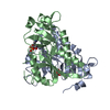
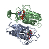
| Structure viewer | Molecule: MolmilJmol/JSmol |
|---|
- Downloads & links
-Download
| PDBx/mmCIF format | 1fsg.cif.gz | 246.4 KB | Display | PDBx/mmCIF format |
|---|---|---|---|---|
| PDB format | pdb1fsg.ent.gz | 198.4 KB | Display | PDB format |
| PDBx/mmJSON format | 1fsg.json.gz | Tree view | PDBx/mmJSON format | |
| Others |  Other downloads Other downloads |
-Validation report
| Arichive directory | https://data.pdbj.org/pub/pdb/validation_reports/fs/1fsgftp://data.pdbj.org/pub/pdb/validation_reports/fs/1fsg | HTTPS FTP |
|---|
-Related structure data
| Related structure data | |
|---|---|
| Similar structure data |


-Links
 PDBj
PDBj
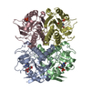
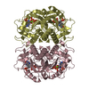
- Assembly
| Deposited unit | 
| ||||||||
|---|---|---|---|---|---|---|---|---|---|
| 1 | 
| ||||||||
| Unit cell |
| ||||||||
| Components on special symmetry positions |
|
-Components
| #1: Protein | Mass: 26705.502 Da / Num. of mol.: 2 Source method: isolated from a genetically manipulated source Source: (gene. exp.)  References: UniProt: Q26997, hypoxanthine phosphoribosyltransferase #2: Chemical | ChemComp-MG /   Mass: 24.305 Da / Num. of mol.: 4 / Source method: obtained synthetically / Formula: Mg Mass: 24.305 Da / Num. of mol.: 4 / Source method: obtained synthetically / Formula: Mg#3: Sugar |   Type: D-saccharide / Mass: 390.070 Da / Num. of mol.: 2 / Source method: obtained synthetically / Formula: C5H13O14P3 Type: D-saccharide / Mass: 390.070 Da / Num. of mol.: 2 / Source method: obtained synthetically / Formula: C5H13O14P3#4: Chemical |   Mass: 150.138 Da / Num. of mol.: 2 / Source method: obtained synthetically / Formula: C6H6N4O Mass: 150.138 Da / Num. of mol.: 2 / Source method: obtained synthetically / Formula: C6H6N4O#5: Water | ChemComp-HOH / |  Mass: 18.015 Da / Num. of mol.: 939 / Source method: isolated from a natural source / Formula: H2O Mass: 18.015 Da / Num. of mol.: 939 / Source method: isolated from a natural source / Formula: H2O |
|---|
-Experimental details
-Experiment
| Experiment | Method: X-RAY DIFFRACTION / Number of used crystals: 1 |
|---|
- Sample preparation
| Crystal | Density Matthews: 2.22 Å3/Da / Density % sol: 45 % | |||||||||||||||||||||||||||||||||||||||||||||||||||||||||||||||
|---|---|---|---|---|---|---|---|---|---|---|---|---|---|---|---|---|---|---|---|---|---|---|---|---|---|---|---|---|---|---|---|---|---|---|---|---|---|---|---|---|---|---|---|---|---|---|---|---|---|---|---|---|---|---|---|---|---|---|---|---|---|---|---|---|
| Crystal grow | Temperature: 277 K / Method: vapor diffusion, hanging drop / pH: 8 Details: THE PROTEIN (20 mg ml-1) WAS MIXED WITH A SOLUTION CONSISTING OF 30% PEG 4000, 100 MM TRIS.HCL (PH 8.0), 200 MM LI2SO4, and 0.5 % BETA-OCTYLGLUCOPYRANOSIDE. THE CRYSTAL WAS GROWN IN THE ...Details: THE PROTEIN (20 mg ml-1) WAS MIXED WITH A SOLUTION CONSISTING OF 30% PEG 4000, 100 MM TRIS.HCL (PH 8.0), 200 MM LI2SO4, and 0.5 % BETA-OCTYLGLUCOPYRANOSIDE. THE CRYSTAL WAS GROWN IN THE PRESENCE OF 2 MM 9-DEAZAGUANINE, 2 MM PRPP AND 10 MM MGCL2 AT 277 K. THE CRYSTAL GREW IN 2 DAYS., VAPOR DIFFUSION, HANGING DROP | |||||||||||||||||||||||||||||||||||||||||||||||||||||||||||||||
| Crystal grow | *PLUS Temperature: 4 ℃Details: drop was mixed with an equal volume of reservoir solution | |||||||||||||||||||||||||||||||||||||||||||||||||||||||||||||||
| Components of the solutions | *PLUS
|
-Data collection
| Diffraction | Mean temperature: 100 K |
|---|---|
| Diffraction source | Source: SYNCHROTRON / Site: SSRL  / Beamline: BL9-1 / Wavelength: 0.783 / Beamline: BL9-1 / Wavelength: 0.783 |
| Detector | Type: MARRESEARCH / Detector: IMAGE PLATE / Date: Mar 1, 1999 / Details: RH-COATED MIRROR |
| Radiation | Monochromator: SI (111) / Protocol: SINGLE WAVELENGTH / Monochromatic (M) / Laue (L): M / Scattering type: x-ray |
| Radiation wavelength | Wavelength: 0.783 Å / Relative weight: 1 |
| Reflection | Resolution: 1.05→35 Å / Num. all: 220504 / Num. obs: 220504 / % possible obs: 99.9 % / Observed criterion σ(F): 0 / Observed criterion σ(I): 0 / Redundancy: 4.2 % / Biso Wilson estimate: 7.435 Å2 / Rmerge(I) obs: 0.072 / Rsym value: 0.072 / Net I/σ(I): 8.4 |
| Reflection shell | Resolution: 1.05→1.08 Å / Redundancy: 3.3 % / Rmerge(I) obs: 0.555 / Mean I/σ(I) obs: 1.8 / Num. unique all: 16129 / Rsym value: 0.555 / % possible all: 99.7 |
| Reflection | *PLUS Num. obs: 220433 / Num. measured all: 915734 |
| Reflection shell | *PLUS % possible obs: 99.7 % |
- Processing
| Software |
| |||||||||||||||||||||||||
|---|---|---|---|---|---|---|---|---|---|---|---|---|---|---|---|---|---|---|---|---|---|---|---|---|---|---|
| Refinement | Method to determine structure: MOLECULAR REPLACEMENT Starting model: UNPUBLISHED T. GONDII HGPRT-GMP COMPLEX STRUCTURE. LOOPS, WATERS AND GMP WERE REMOVED FROM ALL SUBUNITS OF THE MOLECULAR REPLACEMENT MODEL. Resolution: 1.05→35 Å / Cross valid method: THROUGHOUT / σ(F): 0 / σ(I): 0 / Stereochemistry target values: Engh & Huber Details: Refinement with X-PLOR, then REFMAC and ARP alternated with manual rebuilding in O to reduce the free-R to 20.2% (35-1.05A). The model contained at that point alternate conformations, a CIS- ...Details: Refinement with X-PLOR, then REFMAC and ARP alternated with manual rebuilding in O to reduce the free-R to 20.2% (35-1.05A). The model contained at that point alternate conformations, a CIS-peptide bond (Leu78-Lys79), and the active site ligands, all of which were placed in unambiguous electron density. Refinement continued with SHELX-97, with refinement of anisotropic temperature factors. Riding hydrogen atoms were added in the latter stages. NO RESTRAINTS WERE IMPOSED ON THE ATOMS OF 9-DEAZAGUANINE, PHOSPHORIBOSYLPYROPHOSPHATE, THE MAGNESIUM CATIONS AND THEIR SURROUNDING WATER MOLECULES IN THE LATTER STAGES OF THE REFINEMENT.
| |||||||||||||||||||||||||
| Solvent computation | Solvent model: SHELX SWAT | |||||||||||||||||||||||||
| Refinement step | Cycle: LAST / Resolution: 1.05→35 Å
| |||||||||||||||||||||||||
| Refine LS restraints |
| |||||||||||||||||||||||||
| Software | *PLUS Name: SHELXL-97 / Classification: refinement | |||||||||||||||||||||||||
| Refine LS restraints | *PLUS
|
