 Movie
Movie Controller

[English] 日本語
 Yorodumi
- PDB-1hmp: THE CRYSTAL STRUCTURE OF HUMAN HYPOXANTHINE-GUANINE PHOSPHORIBOSY... -
+ Open data

- Basic information
| Entry | Database: PDB / ID: 1hmp | ||||||
|---|---|---|---|---|---|---|---|
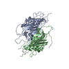
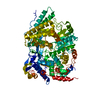
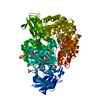
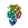
| Title | THE CRYSTAL STRUCTURE OF HUMAN HYPOXANTHINE-GUANINE PHOSPHORIBOSYLTRANSFERASE WITH BOUND GMP | ||||||
 Components Components | HYPOXANTHINE GUANINE PHOSPHORIBOSYL-TRANSFERASE | ||||||
 Keywords Keywords | TRANSFERASE (GLYCOSYLTRANSFERASE) | ||||||
| Function / homology |  Function and homology information Function and homology informationDefective HPRT1 disrupts guanine and hypoxanthine salvage / positive regulation of dopamine metabolic process / GMP catabolic process / adenine metabolic process / sperm annulus / cerebral cortex neuron differentiation / hypoxanthine phosphoribosyltransferase / guanine phosphoribosyltransferase activity / guanine salvage / lymphocyte proliferation ...Defective HPRT1 disrupts guanine and hypoxanthine salvage / positive regulation of dopamine metabolic process / GMP catabolic process / adenine metabolic process / sperm annulus / cerebral cortex neuron differentiation / hypoxanthine phosphoribosyltransferase / guanine phosphoribosyltransferase activity / guanine salvage / lymphocyte proliferation / hypoxanthine metabolic process / hypoxanthine salvage / IMP metabolic process / hypoxanthine phosphoribosyltransferase activity / GMP salvage / Purine salvage / IMP salvage / grooming behavior / AMP salvage / striatum development / dopaminergic neuron differentiation / purine nucleotide biosynthetic process / purine ribonucleoside salvage / Azathioprine ADME / dendrite morphogenesis / central nervous system neuron development / dopamine metabolic process / T cell mediated cytotoxicity / response to amphetamine / sperm end piece / locomotory behavior / sperm principal piece / sperm midpiece / protein homotetramerization / nucleotide binding / magnesium ion binding / extracellular exosome / identical protein binding / cytoplasm / cytosol Similarity search - Function | ||||||
| Biological species |  Homo sapiens (human) Homo sapiens (human) | ||||||
| Method |  X-RAY DIFFRACTION / Resolution: 2.5 Å X-RAY DIFFRACTION / Resolution: 2.5 Å | ||||||
 Authors Authors | Eads, J.C. / Scapin, G. / Xu, Y. / Grubmeyer, C. / Sacchettini, J.C. | ||||||
 Citation Citation | Journal: Cell(Cambridge,Mass.) / Year: 1994 Title: The crystal structure of human hypoxanthine-guanine phosphoribosyltransferase with bound GMP. Authors: Eads, J.C. / Scapin, G. / Xu, Y. / Grubmeyer, C. / Sacchettini, J.C. #1: Journal: Biochemistry / Year: 1994Title: Crystal Structure of Orotate Phosphoribosyltransferase Authors: Scapin, G. / Grubmeyer, C. / Sacchettini, J.C. | ||||||
| History |
|
- Structure visualization
| Structure viewer | Molecule: MolmilJmol/JSmol |
|---|
- Downloads & links
-Download
| PDBx/mmCIF format | 1hmp.cif.gz | 96.7 KB | Display | PDBx/mmCIF format |
|---|---|---|---|---|
| PDB format | pdb1hmp.ent.gz | 74.6 KB | Display | PDB format |
| PDBx/mmJSON format | 1hmp.json.gz | Tree view | PDBx/mmJSON format | |
| Others |  Other downloads Other downloads |
-Validation report
| Arichive directory | https://data.pdbj.org/pub/pdb/validation_reports/hm/1hmpftp://data.pdbj.org/pub/pdb/validation_reports/hm/1hmp | HTTPS FTP |
|---|
-Related structure data
| Similar structure data |
|---|


-Links
 PDBj
PDBj

- Assembly
| Deposited unit | 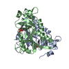
| ||||||||
|---|---|---|---|---|---|---|---|---|---|
| 1 | 
| ||||||||
| Unit cell |
|
-Components
| #1: Protein | Mass: 24481.217 Da / Num. of mol.: 2 Source method: isolated from a genetically manipulated source Source: (gene. exp.) Homo sapiens (human)References: UniProt: P00492, hypoxanthine phosphoribosyltransferase #2: Chemical |   Mass: 363.221 Da / Num. of mol.: 2 / Source method: obtained synthetically / Formula: C10H14N5O8P Mass: 363.221 Da / Num. of mol.: 2 / Source method: obtained synthetically / Formula: C10H14N5O8P#3: Water | ChemComp-HOH / |  Mass: 18.015 Da / Num. of mol.: 85 / Source method: isolated from a natural source / Formula: H2O Mass: 18.015 Da / Num. of mol.: 85 / Source method: isolated from a natural source / Formula: H2ONonpolymer details | THE HET GROUP HAS A CHARGE OF 1- AT PH 5.6. | |
|---|
-Experimental details
-Experiment
| Experiment | Method: X-RAY DIFFRACTION |
|---|
- Sample preparation
| Crystal | Density Matthews: 2.32 Å3/Da / Density % sol: 47.06 % | ||||||||||||||||||||||||||||||||||||||||||
|---|---|---|---|---|---|---|---|---|---|---|---|---|---|---|---|---|---|---|---|---|---|---|---|---|---|---|---|---|---|---|---|---|---|---|---|---|---|---|---|---|---|---|---|
| Crystal grow | *PLUS pH: 5.6 / Method: vapor diffusion, hanging drop | ||||||||||||||||||||||||||||||||||||||||||
| Components of the solutions | *PLUS
|
-Data collection
| Radiation | Scattering type: x-ray |
|---|---|
| Radiation wavelength | Relative weight: 1 |
| Reflection | *PLUS Highest resolution: 2.45 Å / Num. obs: 16428 / % possible obs: 85 % / Rmerge(I) obs: 0.07 |
- Processing
| Software |
| ||||||||||||||||||||||||||||||||||||||||||||||||||||||||||||
|---|---|---|---|---|---|---|---|---|---|---|---|---|---|---|---|---|---|---|---|---|---|---|---|---|---|---|---|---|---|---|---|---|---|---|---|---|---|---|---|---|---|---|---|---|---|---|---|---|---|---|---|---|---|---|---|---|---|---|---|---|---|
| Refinement | Resolution: 2.5→20 Å / σ(F): 1 Details: A LOOP OF RESIDUES 103 - 121 IN BOTH CHAINS A AND B IS POORLY ORDERED. COORDINATES GIVEN FOR THIS REGION RESULT FROM A TENTATIVE FITTING TO POOR ELECTRON DENSITY AND SHOULD BE TREATED WITH ...Details: A LOOP OF RESIDUES 103 - 121 IN BOTH CHAINS A AND B IS POORLY ORDERED. COORDINATES GIVEN FOR THIS REGION RESULT FROM A TENTATIVE FITTING TO POOR ELECTRON DENSITY AND SHOULD BE TREATED WITH CAUTION. FOR THIS LOOP IN THE SECOND MONOMER, RESIDUES 105 - 108 AND 121 ARE MISSING. SOME RESIDUES IN THIS REGION ARE MODELED AS ALANINE RESIDUES.
| ||||||||||||||||||||||||||||||||||||||||||||||||||||||||||||
| Refinement step | Cycle: LAST / Resolution: 2.5→20 Å
| ||||||||||||||||||||||||||||||||||||||||||||||||||||||||||||
| Refine LS restraints |
| ||||||||||||||||||||||||||||||||||||||||||||||||||||||||||||
| Software | *PLUS Name: X-PLOR / Classification: refinement | ||||||||||||||||||||||||||||||||||||||||||||||||||||||||||||
| Refinement | *PLUS Rfactor obs: 0.186 | ||||||||||||||||||||||||||||||||||||||||||||||||||||||||||||
| Solvent computation | *PLUS | ||||||||||||||||||||||||||||||||||||||||||||||||||||||||||||
| Displacement parameters | *PLUS |
