| Entry | Database: PDB / ID: 4pvb
|
|---|
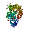
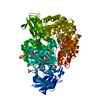
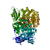
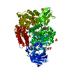
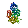
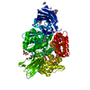
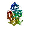
| Title | Crystal structure of Aminopeptidase N in complex with the phosphonic acid analogue of leucine (D-(S)-LeuP) |
|---|
 Components Components | Aminopeptidase N |
|---|
 Keywords Keywords | HYDROLASE / APN / aminopeptidase N / D-(S)-LeuP / Structural Genomics / PSI-Biology / Midwest Center for Structural Genomics / MCSG |
|---|
| Function / homology |  Function and homology information Function and homology information
membrane alanyl aminopeptidase / alanyl aminopeptidase activity / metallopeptidase activity / proteolysis / zinc ion bindingSimilarity search - Function Aminopeptidase N, middle-beta domain / Peptidase M1, alanyl aminopeptidase, C-terminal domain / Peptidase M1, alanyl aminopeptidase / Peptidase M1, alanyl aminopeptidase, C-terminal / Peptidase M1, alanyl aminopeptidase, Ig-like fold / Peptidase M1, alanyl aminopeptidase, C-terminal domain superfamily / Alanyl aminopeptidase, Ig-like domain superfamily / Domain of unknown function (DUF3458) Ig-like fold / Domain of unknown function (DUF3458_C) ARM repeats / Zincin-like fold ...Aminopeptidase N, middle-beta domain / Peptidase M1, alanyl aminopeptidase, C-terminal domain / Peptidase M1, alanyl aminopeptidase / Peptidase M1, alanyl aminopeptidase, C-terminal / Peptidase M1, alanyl aminopeptidase, Ig-like fold / Peptidase M1, alanyl aminopeptidase, C-terminal domain superfamily / Alanyl aminopeptidase, Ig-like domain superfamily / Domain of unknown function (DUF3458) Ig-like fold / Domain of unknown function (DUF3458_C) ARM repeats / Zincin-like fold / Zincin-like - #30 / Zincin-like / tricorn interacting facor f3 domain / Peptidase M1, alanine aminopeptidase/leukotriene A4 hydrolase / Peptidase M1, membrane alanine aminopeptidase / Aminopeptidase N-like , N-terminal domain / Peptidase family M1 domain / Peptidase M1 N-terminal domain / Aminopeptidase N-like , N-terminal domain superfamliy / Neutral Protease Domain 2 / Neutral Protease; domain 2 / Peptidase M4/M1, CTD superfamily / Alpha Horseshoe / Immunoglobulin-like / Sandwich / 2-Layer Sandwich / Orthogonal Bundle / Mainly Beta / Mainly Alpha / Alpha BetaSimilarity search - Domain/homology |
|---|
| Biological species |  Neisseria meningitidis (bacteria) Neisseria meningitidis (bacteria) |
|---|
| Method |  X-RAY DIFFRACTION / SYNCHROTRON / MOLECULAR REPLACEMENT / Resolution: 2.1 Å X-RAY DIFFRACTION / SYNCHROTRON / MOLECULAR REPLACEMENT / Resolution: 2.1 Å |
|---|
 Authors Authors | Nocek, B. / Vassiliou, S. / Berlicki, L. / Mulligan, R. / Mucha, A. / Joachimiak, A. / Midwest Center for Structural Genomics (MCSG) |
|---|
 Citation Citation | Journal: To be Published
Title: Crystal structure of Aminopeptidase N in complex with the phosphonic acid analogue of leucine (D-(S)-LeuP)
Authors: Nocek, B. / Vassiliou, S. / Mulligan, R. / Weglarz-Tomczak, E. / Berlicki, L. / Pawelczak, M. / Mucha, A. / Joachimiak, A. / Midwest Center for Structural Genomics (MCSG) |
|---|
| History | | Deposition | Mar 16, 2014 | Deposition site: RCSB / Processing site: RCSB |
|---|
| Revision 1.0 | Jun 25, 2014 | Provider: repository / Type: Initial release |
|---|
| Revision 1.1 | May 13, 2020 | Group: Derived calculations / Structure summary / Category: audit_author / struct_conn
Item: _audit_author.name / _struct_conn.pdbx_leaving_atom_flag |
|---|
| Revision 1.2 | Sep 20, 2023 | Group: Data collection / Database references ...Data collection / Database references / Derived calculations / Refinement description
Category: chem_comp_atom / chem_comp_bond ...chem_comp_atom / chem_comp_bond / database_2 / pdbx_initial_refinement_model / pdbx_struct_conn_angle / struct_conn / struct_site
Item: _database_2.pdbx_DOI / _database_2.pdbx_database_accession ..._database_2.pdbx_DOI / _database_2.pdbx_database_accession / _pdbx_struct_conn_angle.ptnr1_auth_comp_id / _pdbx_struct_conn_angle.ptnr1_auth_seq_id / _pdbx_struct_conn_angle.ptnr1_label_atom_id / _pdbx_struct_conn_angle.ptnr1_label_comp_id / _pdbx_struct_conn_angle.ptnr1_label_seq_id / _pdbx_struct_conn_angle.ptnr3_auth_comp_id / _pdbx_struct_conn_angle.ptnr3_auth_seq_id / _pdbx_struct_conn_angle.ptnr3_label_atom_id / _pdbx_struct_conn_angle.ptnr3_label_comp_id / _pdbx_struct_conn_angle.ptnr3_label_seq_id / _pdbx_struct_conn_angle.value / _struct_conn.pdbx_dist_value / _struct_conn.ptnr1_auth_comp_id / _struct_conn.ptnr1_auth_seq_id / _struct_conn.ptnr1_label_atom_id / _struct_conn.ptnr1_label_comp_id / _struct_conn.ptnr1_label_seq_id / _struct_site.pdbx_auth_asym_id / _struct_site.pdbx_auth_comp_id / _struct_site.pdbx_auth_seq_id |
|---|
| Revision 1.3 | Dec 6, 2023 | Group: Data collection / Category: chem_comp_atom / chem_comp_bond / Item: _chem_comp_atom.atom_id / _chem_comp_bond.atom_id_2 |
|---|
| Revision 1.4 | Oct 30, 2024 | Group: Structure summary / Category: pdbx_entry_details / pdbx_modification_feature |
|---|
|
|---|
 Movie
Movie Controller

 Yorodumi
 Open data
 Basic information
 Structure visualization
 Downloads & links
 Other downloads


 PDBj
PDBj Assembly


 Mass: 167.143 Da / Num. of mol.: 1 / Source method: obtained synthetically / Formula: C5H14NO3P
 Mass: 65.409 Da / Num. of mol.: 1 / Source method: obtained synthetically / Formula: Zn
 Mass: 96.063 Da / Num. of mol.: 5 / Source method: obtained synthetically / Formula: SO4
 Mass: 94.971 Da / Num. of mol.: 1 / Source method: obtained synthetically / Formula: PO4
 Sample preparation
 / Beamline: 19-ID / Wavelength: 0.9794 Å
 Processing