 Movie
Movie Controller

[English] 日本語
 Yorodumi
- PDB-2al2: Crystal Structure Analysis of Enolase Mg Subunit Complex at pH 8.0 -
+ Open data

- Basic information
| Entry | Database: PDB / ID: 2al2 | ||||||
|---|---|---|---|---|---|---|---|
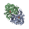
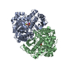
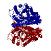
| Title | Crystal Structure Analysis of Enolase Mg Subunit Complex at pH 8.0 | ||||||
 Components Components | (enolase 1) x 2 | ||||||
 Keywords Keywords | LYASE / BETA BARREL | ||||||
| Function / homology |  Function and homology information Function and homology informationGluconeogenesis / regulation of vacuole fusion, non-autophagic / Glycolysis / melatonin binding / phosphopyruvate hydratase / phosphopyruvate hydratase complex / phosphopyruvate hydratase activity / fungal-type vacuole / glycolytic process / magnesium ion binding ...Gluconeogenesis / regulation of vacuole fusion, non-autophagic / Glycolysis / melatonin binding / phosphopyruvate hydratase / phosphopyruvate hydratase complex / phosphopyruvate hydratase activity / fungal-type vacuole / glycolytic process / magnesium ion binding / mitochondrion / plasma membrane / cytoplasm / cytosol Similarity search - Function | ||||||
| Biological species |  | ||||||
| Method |  X-RAY DIFFRACTION / MOLECULAR REPLACEMENT / Resolution: 1.85 Å X-RAY DIFFRACTION / MOLECULAR REPLACEMENT / Resolution: 1.85 Å | ||||||
 Authors Authors | Sims, P.A. / Menefee, A.L. / Larsen, T.M. / Mansoorabadi, S.O. / Reed, G.H. | ||||||
 Citation Citation | Journal: J.Mol.Biol. / Year: 2006 Title: Structure and catalytic properties of an engineered heterodimer of enolase composed of one active and one inactive subunit Authors: Sims, P.A. / Menefee, A.L. / Larsen, T.M. / Mansoorabadi, S.O. / Reed, G.H. | ||||||
| History |
|
- Structure visualization
| Structure viewer | Molecule: MolmilJmol/JSmol |
|---|
- Downloads & links
-Download
| PDBx/mmCIF format | 2al2.cif.gz | 190.2 KB | Display | PDBx/mmCIF format |
|---|---|---|---|---|
| PDB format | pdb2al2.ent.gz | 146.7 KB | Display | PDB format |
| PDBx/mmJSON format | 2al2.json.gz | Tree view | PDBx/mmJSON format | |
| Others |  Other downloads Other downloads |
-Validation report
| Arichive directory | https://data.pdbj.org/pub/pdb/validation_reports/al/2al2ftp://data.pdbj.org/pub/pdb/validation_reports/al/2al2 | HTTPS FTP |
|---|
-Related structure data


-Links
 PDBj
PDBj
- Assembly
| Deposited unit | 
| ||||||||
|---|---|---|---|---|---|---|---|---|---|
| 1 |
| ||||||||
| Unit cell |
|
-Components
-Protein , 2 types, 2 molecules AB
| #1: Protein | Mass: 46674.695 Da / Num. of mol.: 1 Source method: isolated from a genetically manipulated source Details: active form Source: (gene. exp.) Gene: ENO1, ENOA, HSP48 / Plasmid: PetEnol / Production host:  |
|---|---|
| #2: Protein | Mass: 46734.766 Da / Num. of mol.: 1 Source method: isolated from a genetically manipulated source Details: inactive form Source: (gene. exp.) Gene: ENO1, ENOA, HSP48 / Plasmid: PetEnol / Production host: |
-Non-polymers , 6 types, 545 molecules 


| #3: Chemical | ChemComp-MG /  Mass: 24.305 Da / Num. of mol.: 4 / Source method: obtained synthetically / Formula: Mg Mass: 24.305 Da / Num. of mol.: 4 / Source method: obtained synthetically / Formula: Mg#4: Chemical |  Mass: 168.042 Da / Num. of mol.: 2 / Source method: obtained synthetically / Formula: C3H5O6P Mass: 168.042 Da / Num. of mol.: 2 / Source method: obtained synthetically / Formula: C3H5O6P#5: Chemical |  Mass: 186.057 Da / Num. of mol.: 2 / Source method: obtained synthetically / Formula: C3H7O7P Mass: 186.057 Da / Num. of mol.: 2 / Source method: obtained synthetically / Formula: C3H7O7P#6: Chemical | ChemComp-CL / |  Mass: 35.453 Da / Num. of mol.: 1 / Source method: obtained synthetically / Formula: Cl Mass: 35.453 Da / Num. of mol.: 1 / Source method: obtained synthetically / Formula: Cl#7: Chemical | ChemComp-K / |  Mass: 39.098 Da / Num. of mol.: 1 / Source method: obtained synthetically / Formula: K Mass: 39.098 Da / Num. of mol.: 1 / Source method: obtained synthetically / Formula: K#8: Water | ChemComp-HOH / | Mass: 18.015 Da / Num. of mol.: 535 / Source method: isolated from a natural source / Formula: H2O |
|---|
-Details
| Has protein modification | Y |
|---|
-Experimental details
-Experiment
| Experiment | Method: X-RAY DIFFRACTION / Number of used crystals: 1 |
|---|
- Sample preparation
| Crystal | Density Matthews: 2.1 Å3/Da / Density % sol: 42.2 % |
|---|---|
| Crystal grow | Temperature: 293 K / Method: batch / pH: 8 Details: PEG 8000, potasssium chloride, pH 8.0, Batch, temperature 293K |
-Data collection
| Diffraction | Mean temperature: 110 K |
|---|---|
| Diffraction source | Source: ROTATING ANODE / Type: ENRAF-NONIUS FR591 / Wavelength: 1.5418 Å |
| Detector | Type: BRUKER / Detector: CCD / Date: Apr 4, 2005 |
| Radiation | Monochromator: Pt-135 / Protocol: SINGLE WAVELENGTH / Monochromatic (M) / Laue (L): M / Scattering type: x-ray |
| Radiation wavelength | Wavelength: 1.5418 Å / Relative weight: 1 |
| Reflection | Resolution: 1.85→26 Å / Num. all: 67650 / Num. obs: 67650 / % possible obs: 100 % |
- Processing
| Software |
| ||||||||||||||||||||
|---|---|---|---|---|---|---|---|---|---|---|---|---|---|---|---|---|---|---|---|---|---|
| Refinement | Method to determine structure: MOLECULAR REPLACEMENT / Resolution: 1.85→26 Å / σ(F): 0 / Stereochemistry target values: Engh & Huber
| ||||||||||||||||||||
| Refinement step | Cycle: LAST / Resolution: 1.85→26 Å
| ||||||||||||||||||||
| Refine LS restraints |
|
