 Movie
Movie Controller

[English] 日本語
 Yorodumi
- PDB-7enl: MECHANISM OF ENOLASE: THE CRYSTAL STRUCTURE OF ENOLASE-MG2+-PHOSP... -
+ Open data

- Basic information
| Entry | Database: PDB / ID: 7enl | ||||||
|---|---|---|---|---|---|---|---|
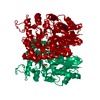
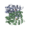
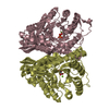
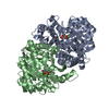
| Title | MECHANISM OF ENOLASE: THE CRYSTAL STRUCTURE OF ENOLASE-MG2+-PHOSPHOGLYCERATE(SLASH) PHOSPHOENOLPYRUVATE COMPLEX AT 2.2-ANGSTROMS RESOLUTION | ||||||
 Components Components | ENOLASE | ||||||
 Keywords Keywords | CARBON-OXYGEN LYASE | ||||||
| Function / homology |  Function and homology information Function and homology informationGluconeogenesis / regulation of vacuole fusion, non-autophagic / Glycolysis / melatonin binding / phosphopyruvate hydratase / phosphopyruvate hydratase complex / phosphopyruvate hydratase activity / fungal-type vacuole / glycolytic process / magnesium ion binding ...Gluconeogenesis / regulation of vacuole fusion, non-autophagic / Glycolysis / melatonin binding / phosphopyruvate hydratase / phosphopyruvate hydratase complex / phosphopyruvate hydratase activity / fungal-type vacuole / glycolytic process / magnesium ion binding / mitochondrion / plasma membrane / cytosol / cytoplasm Similarity search - Function | ||||||
| Biological species |  | ||||||
| Method |  X-RAY DIFFRACTION / Resolution: 2.2 Å X-RAY DIFFRACTION / Resolution: 2.2 Å | ||||||
 Authors Authors | Lebioda, L. / Stec, B. | ||||||
 Citation Citation | Journal: Biochemistry / Year: 1991 Title: Mechanism of enolase: the crystal structure of enolase-Mg2(+)-2-phosphoglycerate/phosphoenolpyruvate complex at 2.2-A resolution. Authors: Lebioda, L. / Stec, B. #1: Journal: Biochemistry / Year: 1991Title: Inhibition of Enolase: The Crystal Structures of Enolase-Ca2+-Phosphoglycerate and Enolase-Zn2+-Phosphoglycolate Complexes at 2.2-Angstroms Resolution Authors: Lebioda, L. / Stec, B. / Brewer, J.M. / Tykarska, E. #2: Journal: J.Mol.Biol. / Year: 1990Title: Refined Structure of Yeast Apo-Enolase at 2.25 Angstroms Resolution Authors: Stec, B. / Lebioda, L. #3: Journal: J.Am.Chem.Soc. / Year: 1989Title: Crystal Structure of Holoenzyme Refined at 1.9 Angstroms Resolution: Trigonal-Bipyramidal Geometry of the Cation Binding Site Authors: Lebioda, L. / Stec, B. #4: Journal: J.Biol.Chem. / Year: 1989Title: The Structure of Yeast Enolase at 2.25-Angstroms Resolution. An 8-Fold Beta+Alpha-Barrel with a Novel Beta Beta Alpha Alpha (Beta Alpha)6 Topology Authors: Lebioda, L. / Stec, B. / Brewer, J.M. #5: Journal: Nature / Year: 1988Title: Crystal Structure of Enolase Indicates that Enolase and Pyruvate Kinase Evolved from a Common Ancestor Authors: Lebioda, L. / Stec, B. #6: Journal: J.Mol.Biol. / Year: 1984Title: Crystallization and Preliminary Crystallographic Data for a Tetragonal Form of Yeast Enolase Authors: Lebioda, L. / Brewer, J.M. | ||||||
| History |
| ||||||
| Remark 700 | SHEET THE SHEET PRESENTED AS *BAR* ON SHEET RECORDS BELOW IS ACTUALLY AN EIGHT-STRANDED BETA-BARREL. ...SHEET THE SHEET PRESENTED AS *BAR* ON SHEET RECORDS BELOW IS ACTUALLY AN EIGHT-STRANDED BETA-BARREL. THIS IS REPRESENTED BY A NINE-STRANDED SHEET IN WHICH THE FIRST AND LAST STRANDS ARE IDENTICAL. THERE IS A SMALL STRAND BONDED TO THE END OF STRAND 1 OF THE BARREL. THIS SMALL STRAND STRAND AND STRAND 1 ARE PRESENTED AS SHEET S1. |
- Structure visualization
| Structure viewer | Molecule: MolmilJmol/JSmol |
|---|
- Downloads & links
-Download
| PDBx/mmCIF format | 7enl.cif.gz | 105.2 KB | Display | PDBx/mmCIF format |
|---|---|---|---|---|
| PDB format | pdb7enl.ent.gz | 79.2 KB | Display | PDB format |
| PDBx/mmJSON format | 7enl.json.gz | Tree view | PDBx/mmJSON format | |
| Others |  Other downloads Other downloads |
-Validation report
| Arichive directory | https://data.pdbj.org/pub/pdb/validation_reports/en/7enlftp://data.pdbj.org/pub/pdb/validation_reports/en/7enl | HTTPS FTP |
|---|
-Related structure data
| Similar structure data |
|---|


-Links
 PDBj
PDBj
- Assembly
| Deposited unit | 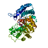
| ||||||||
|---|---|---|---|---|---|---|---|---|---|
| 1 | 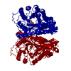
| ||||||||
| Unit cell |
| ||||||||
| Atom site foot note | 1: RESIDUES PRO 143 AND PRO 265 ARE CIS PROLINES. 2: RESIDUES GLY 41 - VAL 42 AND PRO 265 - LYS 269 ARE PROBABLY PARTIALLY DISORDERED. |
-Components
| #1: Protein | Mass: 46690.691 Da / Num. of mol.: 1 Source method: isolated from a genetically manipulated source Source: (gene. exp.) References: UniProt: P00924, phosphopyruvate hydratase |
|---|---|
| #2: Chemical | ChemComp-MG /   Mass: 24.305 Da / Num. of mol.: 1 / Source method: obtained synthetically / Formula: Mg Mass: 24.305 Da / Num. of mol.: 1 / Source method: obtained synthetically / Formula: Mg |
| #3: Chemical | ChemComp-2PG /   Mass: 186.057 Da / Num. of mol.: 1 / Source method: obtained synthetically / Formula: C3H7O7P Mass: 186.057 Da / Num. of mol.: 1 / Source method: obtained synthetically / Formula: C3H7O7P |
| #4: Water | ChemComp-HOH /  Mass: 18.015 Da / Num. of mol.: 347 / Source method: isolated from a natural source / Formula: H2O Mass: 18.015 Da / Num. of mol.: 347 / Source method: isolated from a natural source / Formula: H2O |
-Experimental details
-Experiment
| Experiment | Method: X-RAY DIFFRACTION |
|---|
- Sample preparation
| Crystal | Density Matthews: 2.67 Å3/Da / Density % sol: 53.91 % | ||||||||||||||||||||||||||||||||||||||||||||||||
|---|---|---|---|---|---|---|---|---|---|---|---|---|---|---|---|---|---|---|---|---|---|---|---|---|---|---|---|---|---|---|---|---|---|---|---|---|---|---|---|---|---|---|---|---|---|---|---|---|---|
| Crystal grow | *PLUS pH: 5 / Method: vapor diffusion | ||||||||||||||||||||||||||||||||||||||||||||||||
| Components of the solutions | *PLUS
|
-Data collection
| Reflection | *PLUS Highest resolution: 2.2 Å / Lowest resolution: 8 Å / Num. obs: 21660 / Observed criterion σ(I): 2 / Num. measured all: 16196 / Rmerge(I) obs: 0.075 |
|---|
- Processing
| Software | Name: PROLSQ / Classification: refinement | ||||||||||||||||||||||||||||||||||||||||||||||||||||||||||||||||||||||||||||||||||||
|---|---|---|---|---|---|---|---|---|---|---|---|---|---|---|---|---|---|---|---|---|---|---|---|---|---|---|---|---|---|---|---|---|---|---|---|---|---|---|---|---|---|---|---|---|---|---|---|---|---|---|---|---|---|---|---|---|---|---|---|---|---|---|---|---|---|---|---|---|---|---|---|---|---|---|---|---|---|---|---|---|---|---|---|---|---|
| Refinement | Rfactor obs: 0.169 / Highest resolution: 2.2 Å Details: RESIDUES GLY 41 - VAL 42 AND PRO 265 - LYS 269 ARE PROBABLY PARTIALLY DISORDERED. | ||||||||||||||||||||||||||||||||||||||||||||||||||||||||||||||||||||||||||||||||||||
| Refinement step | Cycle: LAST / Highest resolution: 2.2 Å
| ||||||||||||||||||||||||||||||||||||||||||||||||||||||||||||||||||||||||||||||||||||
| Refine LS restraints |
| ||||||||||||||||||||||||||||||||||||||||||||||||||||||||||||||||||||||||||||||||||||
| Refinement | *PLUS Highest resolution: 2.2 Å / Rfactor all: 0.169 | ||||||||||||||||||||||||||||||||||||||||||||||||||||||||||||||||||||||||||||||||||||
| Solvent computation | *PLUS | ||||||||||||||||||||||||||||||||||||||||||||||||||||||||||||||||||||||||||||||||||||
| Displacement parameters | *PLUS |
