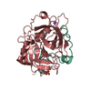
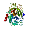
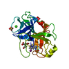
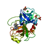
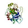
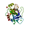
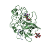
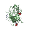
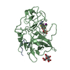
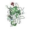
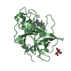
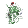
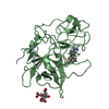
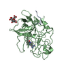
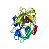
pH: 6.5 詳細: LMW human uPA/A145 was concentrated to 10 mg/ml and incubated in 50 mM HEPES, 5.0 mM NaCl. pH 7.0, 5.0 mM thieno[2,3-b]pyridine-2-carboxamidine for 15 min on ice. The complex was crystallized ...詳細: LMW human uPA/A145 was concentrated to 10 mg/ml and incubated in 50 mM HEPES, 5.0 mM NaCl. pH 7.0, 5.0 mM thieno[2,3-b]pyridine-2-carboxamidine for 15 min on ice. The complex was crystallized by vapor diffusion in hanging drops containing equal volumes of protein-inhibitor solution (0.28 mM uPA/A145, 1.4 mM inhibitor and well solution (20 % 2-propanol, 20 % PEG 4K, 100 mM sodium citrate, pH 6.5) sealed over the well.
プロトコル: SINGLE WAVELENGTH / 単色(M)・ラウエ(L): M / 散乱光タイプ: x-ray
放射波長
波長: 1.5418 Å / 相対比: 1
反射
解像度: 1.44→31.26 Å / Num. all: 23222 / % possible obs: 68 % / Observed criterion σ(I): 0.8 / 冗長度: 1.8 % / Rmerge(I) obs: 0.087 / Net I/σ(I): 7.2
反射 シェル
解像度: 1.65→1.72 Å / Rmerge(I) obs: 0.255 / Mean I/σ(I) obs: 1.5 / % possible all: 32.1
-
解析
ソフトウェア
名称
バージョン
分類
bioteX
(MSC)
データ収集
bioteX
(MSC)
データ削減
X-PLOR
3.1
モデル構築
Quanta
モデル構築
Insight II
モデル構築
X-PLOR
3.1
精密化
bioteX
データスケーリング
X-PLOR
3.1
位相決定
精密化
構造決定の手法: DIFFERENCE FOURIER PLUS REFINEMENT 開始モデル: PDB1LMW 解像度: 1.65→7.5 Å / 交差検証法: X-PLOR / σ(F): 1.9 詳細: Bulk solvent terms included in Fob file created with standard X-PLOR script. Only Leu A9 to Thr_A17 are included for the A-chain. Residues prior and after these residues are not visible ...詳細: Bulk solvent terms included in Fob file created with standard X-PLOR script. Only Leu A9 to Thr_A17 are included for the A-chain. Residues prior and after these residues are not visible (disordered). Residues after Glu_B245 are not visible (disordered). Density for Lys_B243 through Glu_B245 is weak or absent Residues simultaneously refined in two or more conformations are: Ile_B17, Met_B47, Met_B81, Glu_B84, Glu_B86, Leu_B123, Thr_B139, Arg_B166, Gln_B192, Ser_B232, Leu_B235, the inhibitor (246). No energy terms between citrate 1 and 2 are included because they appear to be hydrogen-bonded to one another via an unusually short hydrogen bond between carboxylate groups. Disordered waters are: HOH75 which is close to HOH76; HOH247 which is close to a symmetry-related equivalent of HOH248 which in turn is close to HOH249; HOH104 which is close to HOH105; HOH155 which is close to conformation 1 of Glu_B84; HOH292 which is close to HOH298; HOH373 which is close to HOH374; HOH715 which is close to HOH720 which in turn is close to HOH721; HOH598 WHICH IS CLOSE TO HOH831; HOH70 WHICH IS CLOSE TO HOH364; HOH401 WHICH IS CLOSE TO HOH846; HOH462 WHICH IS CLOSE TO HOH465; HOH451 WHICH IS CLOSE TO HOH719; HOH423 WHICH IS CLOSE TO HOH429; HOH358 WHICH IS CLOSE TO HOH910; HOH30 WHICH IS CLOSE TO A SYMMETRY-RELATED EQUIVALENT OF ITSELF; HOH25 WHICH IS CLOSE TO A SYMMETRY-RELATED EQUIVALENT OF HOH318; HOH38 WHICH IS CLOSE TO A SYMMETRY-RELATED EQUIVALENT OF HOH694; HOH146 WHICH IS CLOSE TO A SYMMETRY-RELATED EQUIVALENT OF HOH375; HOH17 WHICH IS CLOSE TO A SYMMETRY-RELATED EQUIVALENT OF HOH82.
 ムービー
ムービー コントローラー


 データを開く
 基本情報
基本情報 要素
要素 キーワード
キーワード 機能・相同性情報
 Homo sapiens (ヒト)
 X線回折 / DIFFERENCE FOURIER PLUS REFINEMENT / 解像度: 1.65 Å
  データ登録者
 引用
引用 構造の表示
構造の表示 ダウンロードとリンク
 その他のダウンロード
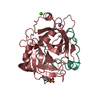


 PDBj
PDBj


 集合体
集合体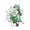
 Pichia pastoris (菌類) / 株 (発現宿主): HISTIDINE DEFICIENT GS115 / 参照: UniProt: P00749, u-plasminogen activator
 分子量: 189.100 Da / 分子数: 3 / 由来タイプ: 合成 / 式: C6H5O7
 分子量: 178.234 Da / 分子数: 1 / 由来タイプ: 合成 / 式: C8H8N3S
 分子量: 18.015 Da / 分子数: 349 / 由来タイプ: 天然 / 式: H2O
 試料調製
試料調製 解析
解析