 Movie
Movie Controller

[English] 日本語
 Yorodumi
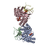
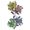
- PDB-1brw: THE CRYSTAL STRUCTURE OF PYRIMIDINE NUCLEOSIDE PHOSPHORYLASE IN A... -
+ Open data

- Basic information
| Entry | Database: PDB / ID: 1brw | ||||||
|---|---|---|---|---|---|---|---|
| Title | THE CRYSTAL STRUCTURE OF PYRIMIDINE NUCLEOSIDE PHOSPHORYLASE IN A CLOSED CONFORMATION | ||||||
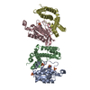
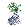
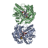
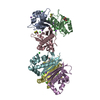
 Components Components | PROTEIN (PYRIMIDINE NUCLEOSIDE PHOSPHORYLASE) | ||||||
 Keywords Keywords | TRANSFERASE / NUCLEOSIDE PHOSPHORYLASE / DOMAIN MOVEMENT | ||||||
| Function / homology |  Function and homology information Function and homology informationpyrimidine-nucleoside phosphorylase / deoxyuridine phosphorylase activity / pyrimidine nucleoside metabolic process / thymidine phosphorylase activity / pyrimidine nucleobase metabolic process / 1,4-alpha-oligoglucan phosphorylase activity / uridine phosphorylase activity / metal ion binding / cytosol Similarity search - Function | ||||||
| Biological species |   Geobacillus stearothermophilus (bacteria) Geobacillus stearothermophilus (bacteria) | ||||||
| Method |  X-RAY DIFFRACTION / SYNCHROTRON / MOLECULAR REPLACEMENT / Resolution: 2.1 Å X-RAY DIFFRACTION / SYNCHROTRON / MOLECULAR REPLACEMENT / Resolution: 2.1 Å | ||||||
 Authors Authors | Pugmire, M.J. / Ealick, S.E. | ||||||
 Citation Citation | Journal: Structure / Year: 1998 Title: The crystal structure of pyrimidine nucleoside phosphorylase in a closed conformation. Authors: Pugmire, M.J. / Ealick, S.E. | ||||||
| History |
|
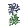
- Structure visualization
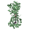
| Structure viewer | Molecule: MolmilJmol/JSmol |
|---|
- Downloads & links
-Download
| PDBx/mmCIF format | 1brw.cif.gz | 173.4 KB | Display | PDBx/mmCIF format |
|---|---|---|---|---|
| PDB format | pdb1brw.ent.gz | 136.4 KB | Display | PDB format |
| PDBx/mmJSON format | 1brw.json.gz | Tree view | PDBx/mmJSON format | |
| Others |  Other downloads Other downloads |
-Validation report
| Arichive directory | https://data.pdbj.org/pub/pdb/validation_reports/br/1brwftp://data.pdbj.org/pub/pdb/validation_reports/br/1brw | HTTPS FTP |
|---|
-Related structure data
| Related structure data |  2tptS S: Starting model for refinement |
|---|---|
| Similar structure data |
-Links
 PDBj
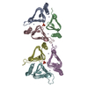
PDBj- Assembly
| Deposited unit | 
| ||||||||
|---|---|---|---|---|---|---|---|---|---|
| 1 |
| ||||||||
| Unit cell |
|
-Components
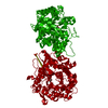
-Protein , 1 types, 2 molecules AB
| #1: Protein | Mass: 46063.184 Da / Num. of mol.: 2 / Source method: isolated from a natural source / Source: (natural) Geobacillus stearothermophilus (bacteria)References: UniProt: P77836, pyrimidine-nucleoside phosphorylase |
|---|
-Non-polymers , 5 types, 117 molecules 


| #2: Chemical |  Mass: 94.971 Da / Num. of mol.: 2 / Source method: obtained synthetically / Formula: PO4 Mass: 94.971 Da / Num. of mol.: 2 / Source method: obtained synthetically / Formula: PO4#3: Chemical |  Mass: 40.078 Da / Num. of mol.: 2 / Source method: obtained synthetically / Formula: Ca Mass: 40.078 Da / Num. of mol.: 2 / Source method: obtained synthetically / Formula: Ca#4: Chemical |  Mass: 195.237 Da / Num. of mol.: 2 / Source method: obtained synthetically / Formula: C6H13NO4S / Comment: pH buffer*YM Mass: 195.237 Da / Num. of mol.: 2 / Source method: obtained synthetically / Formula: C6H13NO4S / Comment: pH buffer*YM#5: Chemical | ChemComp-URA / |  Mass: 112.087 Da / Num. of mol.: 1 / Source method: obtained synthetically / Formula: C4H4N2O2 Mass: 112.087 Da / Num. of mol.: 1 / Source method: obtained synthetically / Formula: C4H4N2O2#6: Water | ChemComp-HOH / | Mass: 18.015 Da / Num. of mol.: 110 / Source method: isolated from a natural source / Formula: H2O |
|---|
-Experimental details
-Experiment
| Experiment | Method: X-RAY DIFFRACTION / Number of used crystals: 1 |
|---|
- Sample preparation
| Crystal | Density Matthews: 2.57 Å3/Da / Density % sol: 52 % | ||||||||||||||||||||||||||||||
|---|---|---|---|---|---|---|---|---|---|---|---|---|---|---|---|---|---|---|---|---|---|---|---|---|---|---|---|---|---|---|---|
| Crystal grow | pH: 6 Details: 0.1 M MES PH 6.0-6.2 25% PEG 6000 PSEUDOURIDINE AT 10X PROTEIN CONCENTRATION | ||||||||||||||||||||||||||||||
| Crystal | *PLUS | ||||||||||||||||||||||||||||||
| Crystal grow | *PLUS pH: 7 / Method: vapor diffusion, hanging drop / Details: Zhou, M., (1998) Acta Cryst., D55, 287. | ||||||||||||||||||||||||||||||
| Components of the solutions | *PLUS
|
-Data collection
| Diffraction | Mean temperature: 108 K |
|---|---|
| Diffraction source | Source: SYNCHROTRON / Site: CHESS  / Beamline: A1 / Wavelength: 0.91 / Beamline: A1 / Wavelength: 0.91 |
| Detector | Type: ADSC / Detector: CCD / Date: Feb 15, 1997 |
| Radiation | Protocol: SINGLE WAVELENGTH / Monochromatic (M) / Laue (L): M / Scattering type: x-ray |
| Radiation wavelength | Wavelength: 0.91 Å / Relative weight: 1 |
| Reflection | Resolution: 2→30 Å / Num. obs: 55716 / % possible obs: 88.4 % / Redundancy: 2.9 % / Rsym value: 0.067 |
| Reflection | *PLUS Num. measured all: 396276 / Rmerge(I) obs: 0.067 |
| Reflection shell | *PLUS Highest resolution: 2 Å / Lowest resolution: 2.07 Å / % possible obs: 41.7 % / Redundancy: 1.6 % / Rmerge(I) obs: 0.232 |
- Processing
| Software |
| ||||||||||||||||||||||||||||||||||||||||||||||||||||||||||||
|---|---|---|---|---|---|---|---|---|---|---|---|---|---|---|---|---|---|---|---|---|---|---|---|---|---|---|---|---|---|---|---|---|---|---|---|---|---|---|---|---|---|---|---|---|---|---|---|---|---|---|---|---|---|---|---|---|---|---|---|---|---|
| Refinement | Method to determine structure: MOLECULAR REPLACEMENT Starting model: 2TPT Resolution: 2.1→30 Å / Data cutoff high absF: 1000000 / Data cutoff low absF: 0.001 / Isotropic thermal model: RESTRAINED / Cross valid method: THROUGHOUT / σ(F): 2
| ||||||||||||||||||||||||||||||||||||||||||||||||||||||||||||
| Displacement parameters | Biso mean: 26.9 Å2 | ||||||||||||||||||||||||||||||||||||||||||||||||||||||||||||
| Refinement step | Cycle: LAST / Resolution: 2.1→30 Å
| ||||||||||||||||||||||||||||||||||||||||||||||||||||||||||||
| Refine LS restraints |
| ||||||||||||||||||||||||||||||||||||||||||||||||||||||||||||
| LS refinement shell | Resolution: 2.1→2.2 Å / Total num. of bins used: 8
| ||||||||||||||||||||||||||||||||||||||||||||||||||||||||||||
| Xplor file | Serial no: 1 / Param file: PARHCSDX.PRO / Topol file: TOPHCSDX.PRO | ||||||||||||||||||||||||||||||||||||||||||||||||||||||||||||
| Software | *PLUS Name: X-PLOR / Version: 3.8 / Classification: refinement | ||||||||||||||||||||||||||||||||||||||||||||||||||||||||||||
| Refinement | *PLUS Highest resolution: 2.1 Å / Lowest resolution: 30 Å / σ(F): 2 / % reflection Rfree: 5 % | ||||||||||||||||||||||||||||||||||||||||||||||||||||||||||||
| Solvent computation | *PLUS | ||||||||||||||||||||||||||||||||||||||||||||||||||||||||||||
| Displacement parameters | *PLUS Biso mean: 26.9 Å2 | ||||||||||||||||||||||||||||||||||||||||||||||||||||||||||||
| Refine LS restraints | *PLUS
| ||||||||||||||||||||||||||||||||||||||||||||||||||||||||||||
| LS refinement shell | *PLUS Highest resolution: 2.1 Å / Lowest resolution: 2.2 Å / % reflection Rfree: 8.6 % |
