 Movie
Movie Controller


+ Open data

- Basic information
| Entry | Database: PDB / ID: 1f7t | ||||||
|---|---|---|---|---|---|---|---|
| Title | HOLO-(ACYL CARRIER PROTEIN) SYNTHASE AT 1.8A | ||||||
 Components Components | HOLO-(ACYL CARRIER PROTEIN) SYNTHASE | ||||||
 Keywords Keywords | TRANSFERASE / 9-strand pseudo beta barrel / LIPID SYNTHESIS | ||||||
| Function / homology |  Function and homology information Function and homology informationholo-[acyl-carrier-protein] synthase / : / holo-[acyl-carrier-protein] synthase activity / fatty acid biosynthetic process / magnesium ion binding / cytosol Similarity search - Function | ||||||
| Biological species |  | ||||||
| Method |  X-RAY DIFFRACTION / SYNCHROTRON / MAD / Resolution: 1.8 Å X-RAY DIFFRACTION / SYNCHROTRON / MAD / Resolution: 1.8 Å | ||||||
 Authors Authors | Parris, K.D. / Lin, L. / Tam, A. / Mathew, R. / Hixon, J. / Stahl, M. / Fritz, C.C. / Seehra, J. / Somers, W.S. | ||||||
 Citation Citation | Journal: Structure Fold.Des. / Year: 2000 Title: Crystal structures of substrate binding to Bacillus subtilis holo-(acyl carrier protein) synthase reveal a novel trimeric arrangement of molecules resulting in three active sites. Authors: Parris, K.D. / Lin, L. / Tam, A. / Mathew, R. / Hixon, J. / Stahl, M. / Fritz, C.C. / Seehra, J. / Somers, W.S. | ||||||
| History |
|
- Structure visualization
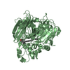
| Structure viewer | Molecule: MolmilJmol/JSmol |
|---|
- Downloads & links
-Download
| PDBx/mmCIF format | 1f7t.cif.gz | 161.9 KB | Display | PDBx/mmCIF format |
|---|---|---|---|---|
| PDB format | pdb1f7t.ent.gz | 128.3 KB | Display | PDB format |
| PDBx/mmJSON format | 1f7t.json.gz | Tree view | PDBx/mmJSON format | |
| Others |  Other downloads Other downloads |
-Validation report
| Arichive directory | https://data.pdbj.org/pub/pdb/validation_reports/f7/1f7tftp://data.pdbj.org/pub/pdb/validation_reports/f7/1f7t | HTTPS FTP |
|---|
-Related structure data


-Links
 PDBj
PDBj


- Assembly
| Deposited unit | 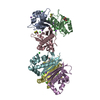
| ||||||||
|---|---|---|---|---|---|---|---|---|---|
| 1 | 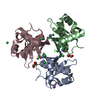
| ||||||||
| 2 | 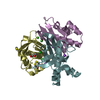
| ||||||||
| 3 | 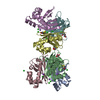
| ||||||||
| Unit cell |
| ||||||||
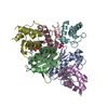
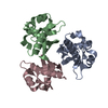
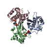
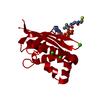
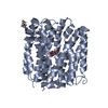
| Details | The biological assembly is a trimer. The asymmetric unit in this structure contains two such trimers, one constructed from chains A, B, and C. The second from chains D, E. F. |
-Components
-Protein , 1 types, 6 molecules ABCDEF
| #1: Protein | Mass: 13691.680 Da / Num. of mol.: 6 / Mutation: Q96P Source method: isolated from a genetically manipulated source Source: (gene. exp.) References: UniProt: P96618, holo-[acyl-carrier-protein] synthase |
|---|
-Non-polymers , 5 types, 503 molecules 


| #2: Chemical | ChemComp-NA /  Mass: 22.990 Da / Num. of mol.: 8 / Source method: obtained synthetically / Formula: Na Mass: 22.990 Da / Num. of mol.: 8 / Source method: obtained synthetically / Formula: Na#3: Chemical | ChemComp-CL /  Mass: 35.453 Da / Num. of mol.: 19 / Source method: obtained synthetically / Formula: Cl Mass: 35.453 Da / Num. of mol.: 19 / Source method: obtained synthetically / Formula: Cl#4: Chemical | ChemComp-DTT /  Mass: 154.251 Da / Num. of mol.: 5 / Source method: obtained synthetically / Formula: C4H10O2S2 Mass: 154.251 Da / Num. of mol.: 5 / Source method: obtained synthetically / Formula: C4H10O2S2#5: Chemical |  Mass: 92.094 Da / Num. of mol.: 2 / Source method: obtained synthetically / Formula: C3H8O3 Mass: 92.094 Da / Num. of mol.: 2 / Source method: obtained synthetically / Formula: C3H8O3#6: Water | ChemComp-HOH / | Mass: 18.015 Da / Num. of mol.: 469 / Source method: isolated from a natural source / Formula: H2O |
|---|
-Experimental details
-Experiment
| Experiment | Method: X-RAY DIFFRACTION / Number of used crystals: 2 |
|---|
- Sample preparation
| Crystal | Density Matthews: 2.8 Å3/Da / Density % sol: 56 % | ||||||||||||||||||||||||
|---|---|---|---|---|---|---|---|---|---|---|---|---|---|---|---|---|---|---|---|---|---|---|---|---|---|
| Crystal grow | Temperature: 291 K / Method: vapor diffusion, hanging drop / pH: 7 Details: Protein: 10mM Sodium Acetate pH 4.4, 2mM Magnesium chloride, 100mM sodium chloride, 5mM Dithiothreitol Well: 2.5M Sodium chloride, 0.1M Tris pH 7.0, 0.2M Magnesium chloride, VAPOR DIFFUSION, ...Details: Protein: 10mM Sodium Acetate pH 4.4, 2mM Magnesium chloride, 100mM sodium chloride, 5mM Dithiothreitol Well: 2.5M Sodium chloride, 0.1M Tris pH 7.0, 0.2M Magnesium chloride, VAPOR DIFFUSION, HANGING DROP, temperature 18K | ||||||||||||||||||||||||
| Crystal grow | *PLUS Method: unknown | ||||||||||||||||||||||||
| Components of the solutions | *PLUS
|
-Data collection
| Diffraction |
| |||||||||||||||
|---|---|---|---|---|---|---|---|---|---|---|---|---|---|---|---|---|
| Diffraction source |
| |||||||||||||||
| Detector |
| |||||||||||||||
| Radiation | Protocol: SINGLE WAVELENGTH / Monochromatic (M) / Laue (L): M / Scattering type: x-ray | |||||||||||||||
| Radiation wavelength |
| |||||||||||||||
| Reflection | Resolution: 1.8→20 Å / Num. all: 87902 / Num. obs: 314385 / % possible obs: 96.4 % / Observed criterion σ(I): -3 / Redundancy: 3.6 % / Biso Wilson estimate: 33.8 Å2 / Rmerge(I) obs: 0.76 / Net I/σ(I): 15.1 | |||||||||||||||
| Reflection shell | Resolution: 1.8→1.83 Å / Redundancy: 4.5 % / Rmerge(I) obs: 0.461 / Num. unique all: 3103 / % possible all: 68.6 | |||||||||||||||
| Reflection | *PLUS Num. obs: 87902 / Num. measured all: 314385 | |||||||||||||||
| Reflection shell | *PLUS % possible obs: 68.6 % / Mean I/σ(I) obs: 1.9 |

- Processing
| Software |
| |||||||||||||||||||||||||
|---|---|---|---|---|---|---|---|---|---|---|---|---|---|---|---|---|---|---|---|---|---|---|---|---|---|---|
| Refinement | Method to determine structure: MAD / Resolution: 1.8→50 Å / σ(F): 0 / Stereochemistry target values: Engh & Huber
| |||||||||||||||||||||||||
| Solvent computation | Solvent model: CNS / Bsol: 57.718 Å2 / ksol: 0.393 e/Å3 | |||||||||||||||||||||||||
| Refinement step | Cycle: LAST / Resolution: 1.8→50 Å
| |||||||||||||||||||||||||
| Refine LS restraints |
| |||||||||||||||||||||||||
| Software | *PLUS Name: CNS / Version: 0.9 / Classification: refinement | |||||||||||||||||||||||||
| Refinement | *PLUS Highest resolution: 1.8 Å / Lowest resolution: 50 Å / σ(F): 0 / % reflection Rfree: 5 % / Rfactor obs: 0.196 | |||||||||||||||||||||||||
| Solvent computation | *PLUS | |||||||||||||||||||||||||
| Displacement parameters | *PLUS | |||||||||||||||||||||||||
| Refine LS restraints | *PLUS Type: c_angle_deg / Dev ideal: 1.3 |
