 Movie
Movie Controller

[English] 日本語
 Yorodumi
- PDB-1f80: HOLO-(ACYL CARRIER PROTEIN) SYNTHASE IN COMPLEX WITH HOLO-(ACYL C... -
+ Open data

- Basic information
| Entry | Database: PDB / ID: 1f80 | ||||||
|---|---|---|---|---|---|---|---|
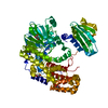
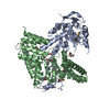
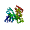
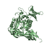
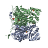
| Title | HOLO-(ACYL CARRIER PROTEIN) SYNTHASE IN COMPLEX WITH HOLO-(ACYL CARRIER PROTEIN) | ||||||
 Components Components |
| ||||||
 Keywords Keywords | TRANSFERASE / 9-strand pseudo beta barrel holo-(acyl carrier protein) acp holo-(acyl carrier protein) synthase acps | ||||||
| Function / homology |  Function and homology information Function and homology informationholo-[acyl-carrier-protein] synthase / : / holo-[acyl-carrier-protein] synthase activity / lipid A biosynthetic process / acyl binding / acyl carrier activity / fatty acid biosynthetic process / magnesium ion binding / membrane / cytosol Similarity search - Function | ||||||
| Biological species |  | ||||||
| Method |  X-RAY DIFFRACTION / MOLECULAR REPLACEMENT / Resolution: 2.3 Å X-RAY DIFFRACTION / MOLECULAR REPLACEMENT / Resolution: 2.3 Å | ||||||
 Authors Authors | Parris, K.D. / Lin, L. / Tam, A. / Mathew, R. / Hixon, J. / Stahl, M. / Fritz, C.C. / Seehra, J. / Somers, W.S. | ||||||
 Citation Citation | Journal: Structure Fold.Des. / Year: 2000 Title: Crystal structures of substrate binding to Bacillus subtilis holo-(acyl carrier protein) synthase reveal a novel trimeric arrangement of molecules resulting in three active sites. Authors: Parris, K.D. / Lin, L. / Tam, A. / Mathew, R. / Hixon, J. / Stahl, M. / Fritz, C.C. / Seehra, J. / Somers, W.S. | ||||||
| History |
|
- Structure visualization
| Structure viewer | Molecule: MolmilJmol/JSmol |
|---|
- Downloads & links
-Download
| PDBx/mmCIF format | 1f80.cif.gz | 123.8 KB | Display | PDBx/mmCIF format |
|---|---|---|---|---|
| PDB format | pdb1f80.ent.gz | 96.6 KB | Display | PDB format |
| PDBx/mmJSON format | 1f80.json.gz | Tree view | PDBx/mmJSON format | |
| Others |  Other downloads Other downloads |
-Validation report
| Arichive directory | https://data.pdbj.org/pub/pdb/validation_reports/f8/1f80ftp://data.pdbj.org/pub/pdb/validation_reports/f8/1f80 | HTTPS FTP |
|---|
-Related structure data
| Related structure data | 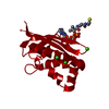 1f7lC  1f7tSC S: Starting model for refinement C: citing same article ( |
|---|---|
| Similar structure data |


-Links
 PDBj
PDBj
- Assembly
| Deposited unit | 
| ||||||||
|---|---|---|---|---|---|---|---|---|---|
| 1 |
| ||||||||
| Unit cell |
| ||||||||
| Details | The biological assembly is a trimer of holo-(acyl protein carrier) synthase molecules. This trimer has three active sites and in each of these active sites, a molecule of holo-(acyl carrier protein) is bound. |
-Components
| #1: Protein | Mass: 13508.407 Da / Num. of mol.: 3 / Mutation: I2A Source method: isolated from a genetically manipulated source Source: (gene. exp.) References: UniProt: P96618, holo-[acyl-carrier-protein] synthase #2: Protein | Mass: 9200.017 Da / Num. of mol.: 3 Source method: isolated from a genetically manipulated source Source: (gene. exp.) #3: Chemical | ChemComp-NA / |   Mass: 22.990 Da / Num. of mol.: 1 / Source method: obtained synthetically / Formula: Na Mass: 22.990 Da / Num. of mol.: 1 / Source method: obtained synthetically / Formula: Na#4: Water | ChemComp-HOH / |  Mass: 18.015 Da / Num. of mol.: 124 / Source method: isolated from a natural source / Formula: H2O Mass: 18.015 Da / Num. of mol.: 124 / Source method: isolated from a natural source / Formula: H2O |
|---|
-Experimental details
-Experiment
| Experiment | Method: X-RAY DIFFRACTION / Number of used crystals: 1 |
|---|
- Sample preparation
| Crystal | Density Matthews: 2.48 Å3/Da / Density % sol: 50 % | ||||||||||||||||||||||||||||||||||||||||||||||||||||||||
|---|---|---|---|---|---|---|---|---|---|---|---|---|---|---|---|---|---|---|---|---|---|---|---|---|---|---|---|---|---|---|---|---|---|---|---|---|---|---|---|---|---|---|---|---|---|---|---|---|---|---|---|---|---|---|---|---|---|
| Crystal grow | Temperature: 291 K / Method: vapor diffusion, hanging drop / pH: 6.4 Details: protein: 50mM Bis-Tris pH 6.4, 100mM sodium chloride, 10mM magnesium chloride, 10mM dithiothreitol Well: 0.15-0.3M potassium formate, 15-23% PEG3350, VAPOR DIFFUSION, HANGING DROP, temperature 18K | ||||||||||||||||||||||||||||||||||||||||||||||||||||||||
| Crystal grow | *PLUS Method: unknown | ||||||||||||||||||||||||||||||||||||||||||||||||||||||||
| Components of the solutions | *PLUS
|
-Data collection
| Diffraction | Mean temperature: 100 K |
|---|---|
| Diffraction source | Source: ROTATING ANODE / Type: RIGAKU RUH2R / Wavelength: 1.54 |
| Detector | Type: RIGAKU RAXIS IV / Detector: IMAGE PLATE / Date: Oct 1, 2000 |
| Radiation | Protocol: SINGLE WAVELENGTH / Monochromatic (M) / Laue (L): M / Scattering type: x-ray |
| Radiation wavelength | Wavelength: 1.54 Å / Relative weight: 1 |
| Reflection | Resolution: 2.3→15 Å / Num. all: 29694 / Num. obs: 270151 / % possible obs: 94.5 % / Observed criterion σ(I): -3 / Redundancy: 9.1 % / Biso Wilson estimate: 47.1 Å2 / Rmerge(I) obs: 0.057 / Net I/σ(I): 26.6 |
| Reflection shell | Resolution: 2.3→2.34 Å / Redundancy: 9.2 % / Rmerge(I) obs: 0.56 / Num. unique all: 1396 / % possible all: 94.5 |
| Reflection | *PLUS Lowest resolution: 15 Å / Num. obs: 29694 / % possible obs: 97.7 % / Num. measured all: 270151 |
| Reflection shell | *PLUS % possible obs: 94.5 % / Rmerge(I) obs: 0.56 / Mean I/σ(I) obs: 3.5 |
- Processing
| Software |
| |||||||||||||||||||||||||
|---|---|---|---|---|---|---|---|---|---|---|---|---|---|---|---|---|---|---|---|---|---|---|---|---|---|---|
| Refinement | Method to determine structure: MOLECULAR REPLACEMENT Starting model: 1F7T Resolution: 2.3→50 Å / σ(F): 0 / Stereochemistry target values: engh and huber
| |||||||||||||||||||||||||
| Solvent computation | Solvent model: CNS / Bsol: 40.302 Å2 / ksol: 0.314 e/Å3 | |||||||||||||||||||||||||
| Refinement step | Cycle: LAST / Resolution: 2.3→50 Å
| |||||||||||||||||||||||||
| Refine LS restraints |
| |||||||||||||||||||||||||
| Software | *PLUS Name: CNS / Version: 0.9 / Classification: refinement | |||||||||||||||||||||||||
| Refinement | *PLUS Highest resolution: 2.3 Å / Lowest resolution: 50 Å / σ(F): 0 / % reflection Rfree: 10 % / Rfactor obs: 0.222 | |||||||||||||||||||||||||
| Solvent computation | *PLUS | |||||||||||||||||||||||||
| Displacement parameters | *PLUS | |||||||||||||||||||||||||
| Refine LS restraints | *PLUS Type: c_angle_deg / Dev ideal: 1.8 |
