 Movie
Movie Controller

[English] 日本語
 Yorodumi
- PDB-6wtf: Structure of radical S-adenosylmethionine methyltransferase, TsrM... -
+ Open data

- Basic information
| Entry | Database: PDB / ID: 6wtf | |||||||||
|---|---|---|---|---|---|---|---|---|---|---|
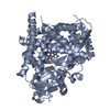
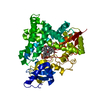
| Title | Structure of radical S-adenosylmethionine methyltransferase, TsrM, from Kitasatospora setae with tryptophan substrate and SAM analog (aza-SAM) bound | |||||||||
 Components Components | Tryptophan-C2-methyltransferase containing B12-binding domain | |||||||||
 Keywords Keywords | TRANSFERASE / Radical SAM / Cobalamin / FeS cluster / methyltransferase | |||||||||
| Function / homology |  Function and homology information Function and homology informationcobalamin binding / catalytic activity / 4 iron, 4 sulfur cluster binding / metal ion binding / cytosol Similarity search - Function | |||||||||
| Biological species |  Kitasatospora setae (bacteria) Kitasatospora setae (bacteria) | |||||||||
| Method |  X-RAY DIFFRACTION / SYNCHROTRON / MOLECULAR REPLACEMENT / Resolution: 2.19 Å X-RAY DIFFRACTION / SYNCHROTRON / MOLECULAR REPLACEMENT / Resolution: 2.19 Å | |||||||||
 Authors Authors | Knox, H.L. / Chen, P.Y.-T. / Drennan, C.L. / Booker, S.J. | |||||||||
| Funding support |  United States, 2items United States, 2items
| |||||||||
 Citation Citation | Journal: Nat.Chem.Biol. / Year: 2021 Title: Structural basis for non-radical catalysis by TsrM, a radical SAM methylase. Authors: Knox, H.L. / Chen, P.Y. / Blaszczyk, A.J. / Mukherjee, A. / Grove, T.L. / Schwalm, E.L. / Wang, B. / Drennan, C.L. / Booker, S.J. | |||||||||
| History |
|
- Structure visualization
| Structure viewer | Molecule: MolmilJmol/JSmol |
|---|
- Downloads & links
-Download
| PDBx/mmCIF format | 6wtf.cif.gz | 237.6 KB | Display | PDBx/mmCIF format |
|---|---|---|---|---|
| PDB format | pdb6wtf.ent.gz | 186.5 KB | Display | PDB format |
| PDBx/mmJSON format | 6wtf.json.gz | Tree view | PDBx/mmJSON format | |
| Others |  Other downloads Other downloads |
-Validation report
| Arichive directory | https://data.pdbj.org/pub/pdb/validation_reports/wt/6wtfftp://data.pdbj.org/pub/pdb/validation_reports/wt/6wtf | HTTPS FTP |
|---|
-Related structure data
| Related structure data |  6wteSC S: Starting model for refinement C: citing same article ( |
|---|---|
| Similar structure data |


-Links
 PDBj
PDBj
- Assembly
| Deposited unit | 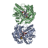
| ||||||||||||||||||||||||||||||||||||||||||||||||||||||||||||||||||||||||||||||||||||||||||
|---|---|---|---|---|---|---|---|---|---|---|---|---|---|---|---|---|---|---|---|---|---|---|---|---|---|---|---|---|---|---|---|---|---|---|---|---|---|---|---|---|---|---|---|---|---|---|---|---|---|---|---|---|---|---|---|---|---|---|---|---|---|---|---|---|---|---|---|---|---|---|---|---|---|---|---|---|---|---|---|---|---|---|---|---|---|---|---|---|---|---|---|
| 1 | 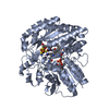
| ||||||||||||||||||||||||||||||||||||||||||||||||||||||||||||||||||||||||||||||||||||||||||
| 2 | 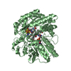
| ||||||||||||||||||||||||||||||||||||||||||||||||||||||||||||||||||||||||||||||||||||||||||
| Unit cell |
| ||||||||||||||||||||||||||||||||||||||||||||||||||||||||||||||||||||||||||||||||||||||||||
| Noncrystallographic symmetry (NCS) | NCS domain:
NCS domain segments: Ens-ID: 1
|