Mass: 366.493 Da / Num. of mol.: 1 / Source method: obtained synthetically / Formula: C24H30O3
-
Experimental details
-
Experiment
Experiment
Method: X-RAY DIFFRACTION
-
Sample preparation
Crystal
Density Matthews: 5.92 Å3/Da / Density % sol: 79.23 %
Crystal grow
Temperature: 277 K / Method: vapor diffusion, sitting drop / pH: 7.4 Details: The enzyme-inhibitor complexes were prepared by the addition from 20mM stock solutions of compound 5 in PEG550 to 18 M (~1mg/ml) of aromatase, to give a final inhibitor concentration of 300 ...Details: The enzyme-inhibitor complexes were prepared by the addition from 20mM stock solutions of compound 5 in PEG550 to 18 M (~1mg/ml) of aromatase, to give a final inhibitor concentration of 300 microM. The mixture was incubated overnight at 4 C in 100mM potassium phosphate buffer pH 7.4 containing 20% glycerol, 20mM dithiothreitol, 0.5 microM ASD and 1mM BDM. The complex was then concentrated to 25-30mg/ml using ultrafiltration. Protein was setup for crystallization using protein to cocktail ratios 2:1 and 3:1. The protein was mixed with reservoir cocktails of 24-30% polyethylene glycol 4000 in 50mM NaCl, 50mM Tris, pH 8.5 and vapor diffused in sealed 24-well sitting drop plates against corresponding reservoir solution, VAPOR DIFFUSION, SITTING DROP, temperature 277K
In the structure databanks used in Yorodumi, some data are registered as the other names, "COVID-19 virus" and "2019-nCoV". Here are the details of the virus and the list of structure data.
Jan 31, 2019. EMDB accession codes are about to change! (news from PDBe EMDB page)
EMDB accession codes are about to change! (news from PDBe EMDB page)
The allocation of 4 digits for EMDB accession codes will soon come to an end. Whilst these codes will remain in use, new EMDB accession codes will include an additional digit and will expand incrementally as the available range of codes is exhausted. The current 4-digit format prefixed with “EMD-” (i.e. EMD-XXXX) will advance to a 5-digit format (i.e. EMD-XXXXX), and so on. It is currently estimated that the 4-digit codes will be depleted around Spring 2019, at which point the 5-digit format will come into force.
The EM Navigator/Yorodumi systems omit the EMD- prefix.
Related info.:Q: What is EMD? / ID/Accession-code notation in Yorodumi/EM Navigator
Yorodumi is a browser for structure data from EMDB, PDB, SASBDB, etc.
This page is also the successor to EM Navigator detail page, and also detail information page/front-end page for Omokage search.
The word "yorodu" (or yorozu) is an old Japanese word meaning "ten thousand". "mi" (miru) is to see.
Related info.:EMDB / PDB / SASBDB / Comparison of 3 databanks / Yorodumi Search / Aug 31, 2016. New EM Navigator & Yorodumi / Yorodumi Papers / Jmol/JSmol / Function and homology information / Changes in new EM Navigator and Yorodumi
 Movie
Movie Controller

 Yorodumi
 Open data
 Basic information
 Components
 Keywords
 Function and homology information
 Homo sapiens (human)
 X-RAY DIFFRACTION /
  Authors
 Citation
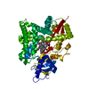
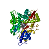
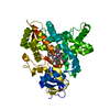
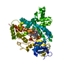
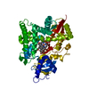
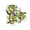
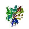
 Structure visualization
 Downloads & links
 Other downloads
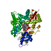


 PDBj
PDBj

 Assembly

 Mass: 616.487 Da / Num. of mol.: 1 / Source method: obtained synthetically / Formula: C34H32FeN4O4
 Mass: 366.493 Da / Num. of mol.: 1 / Source method: obtained synthetically / Formula: C24H30O3
 Sample preparation
 / Beamline: A1 / Wavelength: 0.978 Å
 Processing