[English] 日本語
Yorodumi
- PDB-3eqm: Crystal structure of human placental aromatase cytochrome P450 in... -

+
Open data


ID or keywords:

Loading...

-
Basic information

Entry
Database: PDB / ID: 3eqm
TitleCrystal structure of human placental aromatase cytochrome P450 in complex with androstenedione
ComponentsCytochrome P450 19A1
KeywordsOXIDOREDUCTASE / Human Aromatase / Cytochrome P450 / Membrane Protein / Microsomal / Estrogen / Biosynthesis of Steroid Hormone / Disease mutation / Heme / Iron / Metal-binding / Monooxygenase / Phosphoprotein
Function / homology
Function and homology information


aromatase / Defective CYP19A1 causes AEXS / positive regulation of estradiol secretion / androgen catabolic process / female genitalia development / syncytium formation / estrogen biosynthetic process / negative regulation of chronic inflammatory response / Estrogen biosynthesis / testosterone biosynthetic process ...aromatase / Defective CYP19A1 causes AEXS / positive regulation of estradiol secretion / androgen catabolic process / female genitalia development / syncytium formation / estrogen biosynthetic process / negative regulation of chronic inflammatory response / Estrogen biosynthesis / testosterone biosynthetic process / sterol metabolic process / prostate gland growth / negative regulation of macrophage chemotaxis / mammary gland development / steroid biosynthetic process / steroid hydroxylase activity / oxidoreductase activity, acting on paired donors, with incorporation or reduction of molecular oxygen, reduced flavin or flavoprotein as one donor, and incorporation of one atom of oxygen / aromatase activity / female gonad development / uterus development / Endogenous sterols / oxygen binding / response to estradiol / electron transfer activity / iron ion binding / heme binding / endoplasmic reticulum membrane / endoplasmic reticulum / membrane
Similarity search - Function
Cytochrome P450, E-class, group I / Cytochrome p450 / Cytochrome P450 / Cytochrome P450, conserved site / Cytochrome P450 cysteine heme-iron ligand signature. / Cytochrome P450 / Cytochrome P450 superfamily / Cytochrome P450 / Orthogonal Bundle / Mainly Alpha
Similarity search - Domain/homology
4-ANDROSTENE-3-17-DIONE / PROTOPORPHYRIN IX CONTAINING FE / PHOSPHATE ION / Aromatase
Similarity search - Component
Biological speciesHomo sapiens (human)
MethodX-RAY DIFFRACTION / SYNCHROTRON / MOLECULAR REPLACEMENT / Resolution: 2.9 Å
AuthorsGhosh, D.
CitationJournal: Nature / Year: 2009
Title: Structural basis for androgen specificity and oestrogen synthesis in human aromatase.
Authors: Ghosh, D. / Griswold, J. / Erman, M. / Pangborn, W.
History
DepositionOct 1, 2008Deposition site: RCSB / Processing site: RCSB
Revision 1.0Dec 23, 2008Provider: repository / Type: Initial release
Revision 1.1Jul 13, 2011Group: Version format compliance
Revision 1.2Mar 21, 2012Group: Database references
Revision 1.3Feb 21, 2024Group: Data collection / Database references / Derived calculations
Category: chem_comp_atom / chem_comp_bond ...chem_comp_atom / chem_comp_bond / database_2 / struct_site
Item: _database_2.pdbx_DOI / _database_2.pdbx_database_accession ..._database_2.pdbx_DOI / _database_2.pdbx_database_accession / _struct_site.pdbx_auth_asym_id / _struct_site.pdbx_auth_comp_id / _struct_site.pdbx_auth_seq_id

-
Structure visualization

Structure viewerMolecule:
MolmilJmol/JSmol

Downloads & links

-
Assembly

Deposited unit
A: Cytochrome P450 19A1
hetero molecules


Theoretical massNumber of molelcules
Total (without water)59,0485
Polymers57,9551
Non-polymers1,0934
Water63135
1


  • Idetical with deposited unit
  • defined by author&software
TypeNameSymmetry operationNumber
identity operation1_555x,y,z1
MethodPISA
Unit cell
Length a, b, c (Å)140.208, 140.208, 119.266
Angle α, β, γ (deg.)90.00, 90.00, 120.00
Int Tables number154
Space group name H-MP3221

-
Components

#1: Protein Cytochrome P450 19A1 / Aromatase / CYPXIX / Estrogen synthetase / P-450AROM


Mass: 57954.863 Da / Num. of mol.: 1 / Source method: isolated from a natural source / Source: (natural) Homo sapiens (human) / Organ: placenta / References: UniProt: P11511, unspecific monooxygenase
#2: Chemical ChemComp-HEM / PROTOPORPHYRIN IX CONTAINING FE / HEME / Heme B


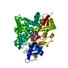
Mass: 616.487 Da / Num. of mol.: 1 / Source method: obtained synthetically / Formula: C34H32FeN4O4
#3: Chemical ChemComp-ASD / 4-ANDROSTENE-3-17-DIONE / Androstenedione


Mass: 286.409 Da / Num. of mol.: 1 / Source method: obtained synthetically / Formula: C19H26O2 / Comment: hormone*YM
#4: Chemical ChemComp-PO4 / PHOSPHATE ION / Phosphate


Mass: 94.971 Da / Num. of mol.: 2 / Source method: obtained synthetically / Formula: PO4
#5: Water ChemComp-HOH / water / Water


Mass: 18.015 Da / Num. of mol.: 35 / Source method: isolated from a natural source / Formula: H2O

-
Experimental details

-
Experiment

ExperimentMethod: X-RAY DIFFRACTION / Number of used crystals: 1

-
Sample preparation

CrystalDensity Matthews: 5.84 Å3/Da / Density % sol: 78.94 %
Crystal growTemperature: 277 K / Method: vapor diffusion / pH: 7.4
Details: Freshly purified aromatase in 100mM potassium phosphate buffer, pH 7.4, containing 20% glycerol, 0.1mM androstenedione, and 1mM n-dodecyl-D-maltopyranoside (BDM) was mixed with reservoir ...Details: Freshly purified aromatase in 100mM potassium phosphate buffer, pH 7.4, containing 20% glycerol, 0.1mM androstenedione, and 1mM n-dodecyl-D-maltopyranoside (BDM) was mixed with reservoir cocktails of 24 to 30% polyethylene glycol 4000 and 0.5M NaCl in 0.05M Tris-HCl buffer pH 8.5 and vapor diffused in sealed 24-well sitting drop plates against corresponding reservoir solution. The purification and crystallization experiments were all conducted at 4 C. Reddish-brown color hexagonal rod-shaped crystals appeared in 7-10 days and continued to grow up to 14-16 days, VAPOR DIFFUSION, temperature 277K

-
Data collection

DiffractionMean temperature: 100 K
Diffraction sourceSource: SYNCHROTRON / Site: APS / Beamline: 19-ID / Wavelength: 0.979 Å
DetectorType: ADSC QUANTUM 315 / Detector: CCD / Date: Nov 28, 2007
RadiationMonochromator: Si / Protocol: SINGLE WAVELENGTH / Monochromatic (M) / Laue (L): M / Scattering type: x-ray
Radiation wavelengthWavelength: 0.979 Å / Relative weight: 1
ReflectionResolution: 2.897→121.27 Å / Num. all: 30371 / Num. obs: 28808 / % possible obs: 99.51 % / Observed criterion σ(F): 0 / Observed criterion σ(I): 0
Reflection shellResolution: 2.897→2.972 Å / Redundancy: 6.1 % / Rmerge(I) obs: 0.067 / Mean I/σ(I) obs: 31.1 / Num. unique all: 30371 / % possible all: 99.6

-
Processing

Software
NameVersionClassification
HKL-3000data collection
AMoREphasing
REFMAC5.2.0019refinement
HKL-3000data reduction
HKL-3000data scaling
RefinementMethod to determine structure: MOLECULAR REPLACEMENT / Resolution: 2.9→37.77 Å / Cor.coef. Fo:Fc: 0.942 / Cor.coef. Fo:Fc free: 0.91 / SU B: 10.2 / SU ML: 0.192 / Cross valid method: THROUGHOUT / σ(F): 0 / ESU R: 0.33 / ESU R Free: 0.261 / Stereochemistry target values: MAXIMUM LIKELIHOOD
RfactorNum. reflection% reflectionSelection details
Rfree0.24439 1532 5 %RANDOM
Rwork0.21372 ---
obs0.21526 28808 99.39 %-
all-30371 --
Solvent computationIon probe radii: 0.8 Å / Shrinkage radii: 0.8 Å / VDW probe radii: 1.2 Å / Solvent model: MASK
Displacement parametersBiso mean: 77.281 Å2
Baniso -1Baniso -2Baniso -3
1-2.89 Å21.44 Å20 Å2
2--2.89 Å20 Å2
3----4.33 Å2
Refinement stepCycle: LAST / Resolution: 2.9→37.77 Å
ProteinNucleic acidLigandSolventTotal
Num. atoms3658 0 74 35 3767
Refine LS restraints
Refine-IDTypeDev idealDev ideal targetNumber
X-RAY DIFFRACTIONr_bond_refined_d0.0090.0223819
X-RAY DIFFRACTIONr_angle_refined_deg1.3212.0165168
X-RAY DIFFRACTIONr_dihedral_angle_1_deg6.145451
X-RAY DIFFRACTIONr_dihedral_angle_2_deg34.6923.576165
X-RAY DIFFRACTIONr_dihedral_angle_3_deg20.15215711
X-RAY DIFFRACTIONr_dihedral_angle_4_deg14.3161525
X-RAY DIFFRACTIONr_chiral_restr0.1020.2564
X-RAY DIFFRACTIONr_gen_planes_refined0.0030.022808
X-RAY DIFFRACTIONr_nbd_refined0.2190.21874
X-RAY DIFFRACTIONr_nbtor_refined0.3110.22643
X-RAY DIFFRACTIONr_xyhbond_nbd_refined0.1550.2122
X-RAY DIFFRACTIONr_symmetry_vdw_refined0.220.224
X-RAY DIFFRACTIONr_symmetry_hbond_refined0.1820.24
X-RAY DIFFRACTIONr_mcbond_it0.4721.52291
X-RAY DIFFRACTIONr_mcangle_it0.8823659
X-RAY DIFFRACTIONr_scbond_it1.00731693
X-RAY DIFFRACTIONr_scangle_it1.7124.51507
LS refinement shellResolution: 2.9→2.972 Å / Total num. of bins used: 20
RfactorNum. reflection% reflection
Rfree0.362 128 -
Rwork0.315 2106 -
obs--99.51 %

+
About Yorodumi

-
News

-
Feb 9, 2022. New format data for meta-information of EMDB entries

New format data for meta-information of EMDB entries

  • Version 3 of the EMDB header file is now the official format.
  • The previous official version 1.9 will be removed from the archive.

Related info.:EMDB header

External links:wwPDB to switch to version 3 of the EMDB data model

-
Aug 12, 2020. Covid-19 info

Covid-19 info

URL: https://pdbj.org/emnavi/covid19.php

New page: Covid-19 featured information page in EM Navigator.

Related info.:Covid-19 info / Mar 5, 2020. Novel coronavirus structure data

+
Mar 5, 2020. Novel coronavirus structure data

Novel coronavirus structure data

Related info.:Yorodumi Speices / Aug 12, 2020. Covid-19 info

External links:COVID-19 featured content - PDBj / Molecule of the Month (242):Coronavirus Proteases

+
Jan 31, 2019. EMDB accession codes are about to change! (news from PDBe EMDB page)

EMDB accession codes are about to change! (news from PDBe EMDB page)

  • The allocation of 4 digits for EMDB accession codes will soon come to an end. Whilst these codes will remain in use, new EMDB accession codes will include an additional digit and will expand incrementally as the available range of codes is exhausted. The current 4-digit format prefixed with “EMD-” (i.e. EMD-XXXX) will advance to a 5-digit format (i.e. EMD-XXXXX), and so on. It is currently estimated that the 4-digit codes will be depleted around Spring 2019, at which point the 5-digit format will come into force.
  • The EM Navigator/Yorodumi systems omit the EMD- prefix.

Related info.:Q: What is EMD? / ID/Accession-code notation in Yorodumi/EM Navigator

External links:EMDB Accession Codes are Changing Soon! / Contact to PDBj

+
Jul 12, 2017. Major update of PDB

Major update of PDB

  • wwPDB released updated PDB data conforming to the new PDBx/mmCIF dictionary.
  • This is a major update changing the version number from 4 to 5, and with Remediation, in which all the entries are updated.
  • In this update, many items about electron microscopy experimental information are reorganized (e.g. em_software).
  • Now, EM Navigator and Yorodumi are based on the updated data.

External links:wwPDB Remediation / Enriched Model Files Conforming to OneDep Data Standards Now Available in the PDB FTP Archive

-
Yorodumi

Thousand views of thousand structures

  • Yorodumi is a browser for structure data from EMDB, PDB, SASBDB, etc.
  • This page is also the successor to EM Navigator detail page, and also detail information page/front-end page for Omokage search.
  • The word "yorodu" (or yorozu) is an old Japanese word meaning "ten thousand". "mi" (miru) is to see.

Related info.:EMDB / PDB / SASBDB / Comparison of 3 databanks / Yorodumi Search / Aug 31, 2016. New EM Navigator & Yorodumi / Yorodumi Papers / Jmol/JSmol / Function and homology information / Changes in new EM Navigator and Yorodumi

Read more