 Movie
Movie Controller

[English] 日本語
 Yorodumi
- PDB-3tio: Crystal structures of yrdA from Escherichia coli, a homologous pr... -
+ Open data

- Basic information
| Entry | Database: PDB / ID: 3tio | ||||||
|---|---|---|---|---|---|---|---|
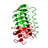
| Title | Crystal structures of yrdA from Escherichia coli, a homologous protein of gamma-class carbonic anhydrase, show possible allosteric conformations | ||||||
 Components Components | Protein YrdA | ||||||
 Keywords Keywords | TRANSFERASE / Carbonic anhydrase (CA) catalyzes / zinc ion binding | ||||||
| Function / homology |  Function and homology information Function and homology informationprotein-containing complex / zinc ion binding / identical protein binding / cytosol Similarity search - Function | ||||||
| Biological species |  | ||||||
| Method |  X-RAY DIFFRACTION / SYNCHROTRON / MOLECULAR REPLACEMENT / Resolution: 1.41 Å X-RAY DIFFRACTION / SYNCHROTRON / MOLECULAR REPLACEMENT / Resolution: 1.41 Å | ||||||
 Authors Authors | Park, H.M. / Choi, J.W. / Lee, J.E. / Jung, C.H. / Kim, B.Y. / Kim, J.S. | ||||||
 Citation Citation | Journal: Acta Crystallogr.,Sect.D / Year: 2012 Title: Structures of the gamma-class carbonic anhydrase homologue YrdA suggest a possible allosteric switch Authors: Park, H.M. / Park, J.H. / Choi, J.W. / Lee, J.E. / Kim, B.Y. / Jung, C.H. / Kim, J.S. | ||||||
| History |
|
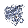
- Structure visualization
| Structure viewer | Molecule: MolmilJmol/JSmol |
|---|
- Downloads & links
-Download
| PDBx/mmCIF format | 3tio.cif.gz | 238.1 KB | Display | PDBx/mmCIF format |
|---|---|---|---|---|
| PDB format | pdb3tio.ent.gz | 189.1 KB | Display | PDB format |
| PDBx/mmJSON format | 3tio.json.gz | Tree view | PDBx/mmJSON format | |
| Others |  Other downloads Other downloads |
-Validation report
| Arichive directory | https://data.pdbj.org/pub/pdb/validation_reports/ti/3tioftp://data.pdbj.org/pub/pdb/validation_reports/ti/3tio | HTTPS FTP |
|---|
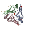
-Related structure data
| Related structure data |  3tisC 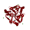 1v3wS S: Starting model for refinement C: citing same article ( |
|---|---|
| Similar structure data |


-Links
 PDBj
PDBj


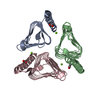
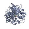
- Assembly
| Deposited unit | 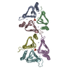
| ||||||||
|---|---|---|---|---|---|---|---|---|---|
| 1 | 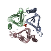
| ||||||||
| 2 | 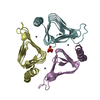
| ||||||||
| Unit cell |
|
-Components
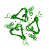
| #1: Protein | Mass: 20116.754 Da / Num. of mol.: 6 / Mutation: R6H Source method: isolated from a genetically manipulated source Source: (gene. exp.) #2: Chemical | ChemComp-ZN /   Mass: 65.409 Da / Num. of mol.: 6 / Source method: obtained synthetically / Formula: Zn Mass: 65.409 Da / Num. of mol.: 6 / Source method: obtained synthetically / Formula: Zn#3: Chemical | ChemComp-PO4 /   Mass: 94.971 Da / Num. of mol.: 4 / Source method: obtained synthetically / Formula: PO4 Mass: 94.971 Da / Num. of mol.: 4 / Source method: obtained synthetically / Formula: PO4#4: Water | ChemComp-HOH / |  Mass: 18.015 Da / Num. of mol.: 1216 / Source method: isolated from a natural source / Formula: H2O Mass: 18.015 Da / Num. of mol.: 1216 / Source method: isolated from a natural source / Formula: H2O |
|---|
-Experimental details
-Experiment
| Experiment | Method: X-RAY DIFFRACTION / Number of used crystals: 1 |
|---|
- Sample preparation
| Crystal | Density Matthews: 1.91 Å3/Da / Density % sol: 35.6 % |
|---|---|
| Crystal grow | Temperature: 295 K / Method: evaporation / pH: 4.6 Details: 20 % (w/v) polyethyleneglycol Monomethyl Ether 2000, 0.15M (NH4)2SO4, 0.1M Sodium Acetate, pH 4.6, EVAPORATION, temperature 295K |
-Data collection
| Diffraction | Mean temperature: 100 K |
|---|---|
| Diffraction source | Source: SYNCHROTRON / Site: Photon Factory  / Beamline: BL-17A / Wavelength: 1 Å / Beamline: BL-17A / Wavelength: 1 Å |
| Detector | Type: ADSC QUANTUM 315r / Detector: CCD / Date: Feb 22, 2011 |
| Radiation | Protocol: SINGLE WAVELENGTH / Monochromatic (M) / Laue (L): M / Scattering type: x-ray |
| Radiation wavelength | Wavelength: 1 Å / Relative weight: 1 |
| Reflection | Resolution: 1.41→50 Å / Num. obs: 173327 / % possible obs: 99.3 % / Observed criterion σ(F): -3 / Observed criterion σ(I): -3 |
| Reflection shell | Resolution: 1.41→1.43 Å / % possible all: 99.3 |
- Processing
| Software |
| ||||||||||||||||||
|---|---|---|---|---|---|---|---|---|---|---|---|---|---|---|---|---|---|---|---|
| Refinement | Method to determine structure: MOLECULAR REPLACEMENT Starting model: 1V3W Resolution: 1.41→50 Å / σ(F): -3
| ||||||||||||||||||
| Refinement step | Cycle: LAST / Resolution: 1.41→50 Å
| ||||||||||||||||||
| Refine LS restraints |
|
