 Movie
Movie Controller


+ Open data

- Basic information
| Entry | Database: PDB / ID: 3mdv | ||||||
|---|---|---|---|---|---|---|---|
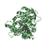
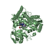
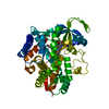
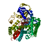
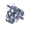
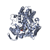
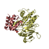
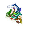
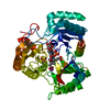
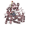
| Title | Clotrimazole complex of Cytochrome P450 46A1 | ||||||
 Components Components | Cholesterol 24-hydroxylase | ||||||
 Keywords Keywords | OXIDOREDUCTASE / CYP46A1 / P450 46A1 / P450 / CLOTRIMAZOLE / MONOOXYGENASE / METABOLIC ENZYME / HEME / Cholesterol metabolism / Endoplasmic reticulum / Iron / Lipid metabolism / Membrane / Metal-binding / Microsome / NADP / Steroid metabolism / Transmembrane | ||||||
| Function / homology |  Function and homology information Function and homology informationcholesterol 24-hydroxylase / cholesterol 24-hydroxylase activity / protein localization to membrane raft / testosterone 16-beta-hydroxylase activity / Synthesis of bile acids and bile salts via 24-hydroxycholesterol / bile acid biosynthetic process / testosterone 6-beta-hydroxylase activity / progesterone metabolic process / sterol metabolic process / steroid hydroxylase activity ...cholesterol 24-hydroxylase / cholesterol 24-hydroxylase activity / protein localization to membrane raft / testosterone 16-beta-hydroxylase activity / Synthesis of bile acids and bile salts via 24-hydroxycholesterol / bile acid biosynthetic process / testosterone 6-beta-hydroxylase activity / progesterone metabolic process / sterol metabolic process / steroid hydroxylase activity / cholesterol catabolic process / regulation of long-term synaptic potentiation / Endogenous sterols / xenobiotic metabolic process / nervous system development / presynapse / postsynapse / iron ion binding / heme binding / dendrite / endoplasmic reticulum membrane / endoplasmic reticulum Similarity search - Function | ||||||
| Biological species |  Homo sapiens (human) Homo sapiens (human) | ||||||
| Method |  X-RAY DIFFRACTION / SYNCHROTRON / MOLECULAR REPLACEMENT / Resolution: 2.4 Å X-RAY DIFFRACTION / SYNCHROTRON / MOLECULAR REPLACEMENT / Resolution: 2.4 Å | ||||||
 Authors Authors | Mast, N. / Charvet, C. / Pikuleva, I. / Stout, C.D. | ||||||
 Citation Citation | Journal: J.Biol.Chem. / Year: 2010 Title: Structural basis of drug binding to CYP46A1, an enzyme that controls cholesterol turnover in the brain. Authors: Mast, N. / Charvet, C. / Pikuleva, I.A. / Stout, C.D. #1: Journal: Proc.Natl.Acad.Sci.USA / Year: 2008Title: Crystal structures of substrate-bound and substrate-free cytochrome P450 46A1, the principal cholesterol hydroxylase in the brain. Authors: Mast, N. / White, M.A. / Bjorkhem, I. / Johnson, E.F. / Stout, C.D. / Pikuleva, I.A. | ||||||
| History |
|
- Structure visualization
| Structure viewer | Molecule: MolmilJmol/JSmol |
|---|
- Downloads & links
-Download
| PDBx/mmCIF format | 3mdv.cif.gz | 197.9 KB | Display | PDBx/mmCIF format |
|---|---|---|---|---|
| PDB format | pdb3mdv.ent.gz | 155.7 KB | Display | PDB format |
| PDBx/mmJSON format | 3mdv.json.gz | Tree view | PDBx/mmJSON format | |
| Others |  Other downloads Other downloads |
-Validation report
| Arichive directory | https://data.pdbj.org/pub/pdb/validation_reports/md/3mdvftp://data.pdbj.org/pub/pdb/validation_reports/md/3mdv | HTTPS FTP |
|---|
-Related structure data
| Related structure data |  3mdmC  3mdrC  3mdtC  2q9fS S: Starting model for refinement C: citing same article ( |
|---|---|
| Similar structure data |

-Links
 PDBj
PDBj


- Assembly
| Deposited unit | 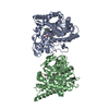
| ||||||||||||||||||
|---|---|---|---|---|---|---|---|---|---|---|---|---|---|---|---|---|---|---|---|
| 1 | 
| ||||||||||||||||||
| 2 | 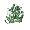
| ||||||||||||||||||
| Unit cell |
| ||||||||||||||||||
| Components on special symmetry positions |
| ||||||||||||||||||
| Noncrystallographic symmetry (NCS) | NCS domain:
NCS domain segments: Component-ID: 1 / Ens-ID: 1 / Beg auth comp-ID: VAL / Beg label comp-ID: VAL / End auth comp-ID: ARG / End label comp-ID: ARG / Refine code: 3 / Auth seq-ID: 59 - 489 / Label seq-ID: 11 - 441
|
-Components
| #1: Protein | Mass: 52125.086 Da / Num. of mol.: 2 / Fragment: UNP residues 51-500 Source method: isolated from a genetically manipulated source Source: (gene. exp.) Homo sapiens (human) / Gene: CYP46A1, CYP46 / Plasmid: PUC18 / Production host:  #2: Chemical |   Mass: 616.487 Da / Num. of mol.: 2 / Source method: obtained synthetically / Formula: C34H32FeN4O4 Mass: 616.487 Da / Num. of mol.: 2 / Source method: obtained synthetically / Formula: C34H32FeN4O4#3: Chemical |   Mass: 344.837 Da / Num. of mol.: 2 / Source method: obtained synthetically / Formula: C22H17ClN2 Mass: 344.837 Da / Num. of mol.: 2 / Source method: obtained synthetically / Formula: C22H17ClN2#4: Water | ChemComp-HOH / |  Mass: 18.015 Da / Num. of mol.: 484 / Source method: isolated from a natural source / Formula: H2O Mass: 18.015 Da / Num. of mol.: 484 / Source method: isolated from a natural source / Formula: H2O |
|---|
-Experimental details
-Experiment
| Experiment | Method: X-RAY DIFFRACTION / Number of used crystals: 1 |
|---|
- Sample preparation
| Crystal | Density Matthews: 2.6 Å3/Da / Density % sol: 52.73 % |
|---|---|
| Crystal grow | Temperature: 291 K / Method: vapor diffusion, sitting drop / pH: 6.4 Details: 14% PEG 8000, 20% glycerol, 50 mM KPi, pH 6.4, VAPOR DIFFUSION, SITTING DROP, temperature 291K |
-Data collection
| Diffraction | Mean temperature: 100 K | |||||||||||||||
|---|---|---|---|---|---|---|---|---|---|---|---|---|---|---|---|---|
| Diffraction source | Source: SYNCHROTRON / Site: SSRL  / Beamline: BL7-1 / Wavelength: 0.979 Å / Beamline: BL7-1 / Wavelength: 0.979 Å | |||||||||||||||
| Detector | Type: ADSC QUANTUM 315r / Detector: CCD / Date: Jul 31, 2009 / Details: Rh coated flat mirror | |||||||||||||||
| Radiation | Monochromator: side scattering I-beam bent single crystal, asymmetric cut 4.9650 deg Protocol: SINGLE WAVELENGTH / Scattering type: x-ray | |||||||||||||||
| Radiation wavelength | Wavelength: 0.979 Å / Relative weight: 1 | |||||||||||||||
| Reflection twin |
| |||||||||||||||
| Reflection | Resolution: 2.4→93.074 Å / Num. all: 39099 / Num. obs: 39099 / % possible obs: 95.3 % / Observed criterion σ(F): 0 / Observed criterion σ(I): 0 / Redundancy: 4.9 % / Biso Wilson estimate: 42.7 Å2 / Rmerge(I) obs: 0.104 / Rsym value: 0.104 / Net I/σ(I): 4.6 | |||||||||||||||
| Reflection shell | Resolution: 2.4→2.53 Å / Redundancy: 4.9 % / Rmerge(I) obs: 0.396 / Mean I/σ(I) obs: 1.9 / Num. unique all: 5847 / Rsym value: 0.396 / % possible all: 97.2 |
- Processing
| Software |
| |||||||||||||||||||||||||||||||||||||||||||||||||||||||||||||||||
|---|---|---|---|---|---|---|---|---|---|---|---|---|---|---|---|---|---|---|---|---|---|---|---|---|---|---|---|---|---|---|---|---|---|---|---|---|---|---|---|---|---|---|---|---|---|---|---|---|---|---|---|---|---|---|---|---|---|---|---|---|---|---|---|---|---|---|
| Refinement | Method to determine structure: MOLECULAR REPLACEMENT Starting model: PDB ENTRY 2Q9F Resolution: 2.4→30 Å / Cor.coef. Fo:Fc: 0.914 / Cor.coef. Fo:Fc free: 0.909 / Occupancy max: 1 / Occupancy min: 0.5 / SU B: 6.573 / SU ML: 0.153 / Isotropic thermal model: ISOTROPIC / Cross valid method: THROUGHOUT / σ(F): 0 / ESU R: 0.119 / ESU R Free: 0.055 / Stereochemistry target values: MAXIMUM LIKELIHOOD Details: HYDROGENS HAVE BEEN ADDED IN THE RIDING POSITIONS U VALUES: REFINED INDIVIDUALLY TWIN DOMAINS: H,K,L TWIN FRACTION 0.459 -H,K,-L TWIN FRACTION 0.541
| |||||||||||||||||||||||||||||||||||||||||||||||||||||||||||||||||
| Solvent computation | Ion probe radii: 0.8 Å / Shrinkage radii: 0.8 Å / VDW probe radii: 1.4 Å / Solvent model: MASK | |||||||||||||||||||||||||||||||||||||||||||||||||||||||||||||||||
| Displacement parameters | Biso max: 71.73 Å2 / Biso mean: 32.556 Å2 / Biso min: 2.52 Å2
| |||||||||||||||||||||||||||||||||||||||||||||||||||||||||||||||||
| Refinement step | Cycle: LAST / Resolution: 2.4→30 Å
| |||||||||||||||||||||||||||||||||||||||||||||||||||||||||||||||||
| Refine LS restraints |
| |||||||||||||||||||||||||||||||||||||||||||||||||||||||||||||||||
| Refine LS restraints NCS | Dom-ID: 1 / Auth asym-ID: B / Ens-ID: 1 / Refine-ID: X-RAY DIFFRACTION
| |||||||||||||||||||||||||||||||||||||||||||||||||||||||||||||||||
| LS refinement shell | Resolution: 2.4→2.462 Å / Total num. of bins used: 20
|
