 Movie
Movie Controller


+ Open data

- Basic information
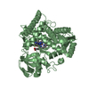
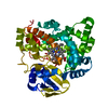
| Entry | Database: PDB / ID: 2q9g | ||||||
|---|---|---|---|---|---|---|---|
| Title | Crystal structure of human cytochrome P450 46A1 | ||||||
 Components Components | Cytochrome P450 46A1 | ||||||
 Keywords Keywords | OXIDOREDUCTASE / CYP46A1 / P450 46A1 / P450 / MONOOXYGENASE / CHOLESTEROL METABOLIC ENZYME / HEME | ||||||
| Function / homology |  Function and homology information Function and homology informationcholesterol 24-hydroxylase / cholesterol 24-hydroxylase activity / protein localization to membrane raft / testosterone 16-beta-hydroxylase activity / Synthesis of bile acids and bile salts via 24-hydroxycholesterol / bile acid biosynthetic process / testosterone 6-beta-hydroxylase activity / progesterone metabolic process / sterol metabolic process / steroid hydroxylase activity ...cholesterol 24-hydroxylase / cholesterol 24-hydroxylase activity / protein localization to membrane raft / testosterone 16-beta-hydroxylase activity / Synthesis of bile acids and bile salts via 24-hydroxycholesterol / bile acid biosynthetic process / testosterone 6-beta-hydroxylase activity / progesterone metabolic process / sterol metabolic process / steroid hydroxylase activity / cholesterol catabolic process / regulation of long-term synaptic potentiation / Endogenous sterols / xenobiotic metabolic process / nervous system development / presynapse / postsynapse / iron ion binding / heme binding / dendrite / endoplasmic reticulum membrane / endoplasmic reticulum Similarity search - Function | ||||||
| Biological species |  Homo sapiens (human) Homo sapiens (human) | ||||||
| Method |  X-RAY DIFFRACTION / SYNCHROTRON / MOLECULAR REPLACEMENT / Resolution: 2.4 Å X-RAY DIFFRACTION / SYNCHROTRON / MOLECULAR REPLACEMENT / Resolution: 2.4 Å | ||||||
 Authors Authors | White, M.A. / Mast, N.V. / Johnson, E.F. / Stout, C.D. / Pikuleva, I.A. | ||||||
 Citation Citation | Journal: Proc.Natl.Acad.Sci.Usa / Year: 2008 Title: Crystal structures of substrate-bound and substrate-free cytochrome P450 46A1, the principal cholesterol hydroxylase in the brain. Authors: Mast, N. / White, M.A. / Bjorkhem, I. / Johnson, E.F. / Stout, C.D. / Pikuleva, I.A. #1: Journal: Acta Crystallogr.,Sect.D / Year: 2008 Title: Use of complementary cation and anion heavy-atom salt derivatives to solve the structure of cytochrome P450 46A1. Authors: White, M.A. / Mast, N. / Bjorkhem, I. / Johnson, E.F. / Stout, C.D. / Pikuleva, I.A. | ||||||
| History |
|
- Structure visualization
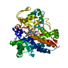
| Structure viewer | Molecule: MolmilJmol/JSmol |
|---|
- Downloads & links
-Download
| PDBx/mmCIF format | 2q9g.cif.gz | 102.9 KB | Display | PDBx/mmCIF format |
|---|---|---|---|---|
| PDB format | pdb2q9g.ent.gz | 76.6 KB | Display | PDB format |
| PDBx/mmJSON format | 2q9g.json.gz | Tree view | PDBx/mmJSON format | |
| Others |  Other downloads Other downloads |
-Validation report
| Arichive directory | https://data.pdbj.org/pub/pdb/validation_reports/q9/2q9gftp://data.pdbj.org/pub/pdb/validation_reports/q9/2q9g | HTTPS FTP |
|---|
-Related structure data
| Related structure data |  2q9fSC S: Starting model for refinement C: citing same article ( |
|---|---|
| Similar structure data |


-Links
 PDBj
PDBj


- Assembly
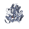
| Deposited unit | 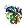
| ||||||||
|---|---|---|---|---|---|---|---|---|---|
| 1 |
| ||||||||
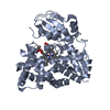
| Unit cell |
|
-Components
| #1: Protein | Mass: 52125.086 Da / Num. of mol.: 1 / Fragment: RESIDUES 51-500 Source method: isolated from a genetically manipulated source Source: (gene. exp.) Homo sapiens (human) / Gene: CYP46A1, CYP46 / Plasmid: PUC18 / Production host:  | ||
|---|---|---|---|
| #2: Chemical | ChemComp-HEM /   Mass: 616.487 Da / Num. of mol.: 1 / Source method: obtained synthetically / Formula: C34H32FeN4O4 Mass: 616.487 Da / Num. of mol.: 1 / Source method: obtained synthetically / Formula: C34H32FeN4O4 | ||
| #3: Chemical |   Mass: 92.094 Da / Num. of mol.: 2 / Source method: obtained synthetically / Formula: C3H8O3 Mass: 92.094 Da / Num. of mol.: 2 / Source method: obtained synthetically / Formula: C3H8O3#4: Water | ChemComp-HOH / |  Mass: 18.015 Da / Num. of mol.: 25 / Source method: isolated from a natural source / Formula: H2O Mass: 18.015 Da / Num. of mol.: 25 / Source method: isolated from a natural source / Formula: H2O |
-Experimental details
-Experiment
| Experiment | Method: X-RAY DIFFRACTION / Number of used crystals: 1 |
|---|
- Sample preparation
| Crystal | Density Matthews: 2.54 Å3/Da / Density % sol: 51.65 % |
|---|---|
| Crystal grow | Temperature: 291 K / Method: vapor diffusion, sitting drop / pH: 4.6 Details: PEG8000, Potassium Phosphate, Glycerol, NaCl, PH 4.6, VAPOR DIFFUSION, HANGING DROP, TEMPERATURE 291K, VAPOR DIFFUSION, SITTING DROP |
-Data collection
| Diffraction | Mean temperature: 100 K |
|---|---|
| Diffraction source | Source: SYNCHROTRON / Site: SSRL  / Beamline: BL11-1 / Wavelength: 0.97945 Å / Beamline: BL11-1 / Wavelength: 0.97945 Å |
| Detector | Type: ADSC QUANTUM 315 / Detector: CCD / Date: Jan 30, 2007 Details: FLAT MIRROR (VERTICAL FOCUSING), SINGLE CRYSTAL SI(111) BENT MONOCHROMATOR (HO RIZONTAL FOCUSING) |
| Radiation | Monochromator: SIDE SCATTERING BENT CUBE- ROOT I-BEAM SINGLE CRYSTAL, ASYMMETRIC CUT 4.965 DEGS Protocol: SINGLE WAVELENGTH / Monochromatic (M) / Laue (L): M / Scattering type: x-ray |
| Radiation wavelength | Wavelength: 0.97945 Å / Relative weight: 1 |
| Reflection | Resolution: 2.4→92.45 Å / Num. all: 21106 / Num. obs: 21106 / % possible obs: 99.8 % / Observed criterion σ(F): 0 / Observed criterion σ(I): 0 / Redundancy: 7 % / Biso Wilson estimate: 48 Å2 / Rmerge(I) obs: 0.098 / Rsym value: 0.098 / Net I/σ(I): 34.6 |
| Reflection shell | Resolution: 2.4→2.46 Å / Redundancy: 7.2 % / Rmerge(I) obs: 0.9 / Mean I/σ(I) obs: 1.8 / Rsym value: 0.9 / % possible all: 100 |
- Processing
| Software |
| ||||||||||||||||||||||||||||||||||||||||||||||||||||||||||||
|---|---|---|---|---|---|---|---|---|---|---|---|---|---|---|---|---|---|---|---|---|---|---|---|---|---|---|---|---|---|---|---|---|---|---|---|---|---|---|---|---|---|---|---|---|---|---|---|---|---|---|---|---|---|---|---|---|---|---|---|---|---|
| Refinement | Method to determine structure: MOLECULAR REPLACEMENT Starting model: CYP46A1-C3S, PDB ENTRY 2q9f Resolution: 2.4→46.21 Å / Rfactor Rfree error: 0.008 / Data cutoff high absF: 4160886 / Data cutoff low absF: 0 / Isotropic thermal model: variable / Cross valid method: THROUGHOUT / σ(F): 0 / σ(I): 0 / Stereochemistry target values: Engh & Huber Details: Refmac5 and TLS used in intermediate refinement steps. TLS not used in CNS refinement. TLS groups determined using the TLSMD server. TLSMD REF: J Painter & E A Merritt (2006) J. Appl. Cryst. ...Details: Refmac5 and TLS used in intermediate refinement steps. TLS not used in CNS refinement. TLS groups determined using the TLSMD server. TLSMD REF: J Painter & E A Merritt (2006) J. Appl. Cryst. 39, 109-111. PMB used in all CNS refinements. PMB BOND target set to 0.011. PMB Variable Sigma-B used default parameters. PMB Hydrogen bond NOE restraints used: D=2.90, 0.4, 0.7, K=75, PMB Hydrogen bond NOE restraints used: Number of NOEs=272. PMB Local scale reject set to 35 sigma, PMB Local Scale rejected 103 reflections of 21096. PMB Local Scale (h,k,l) box sizes = +/- 4 3 5, PMB Local Scale maximum scale used (localscale method): 32.710, PMB Local Scale minimum scale used (localscale method): 1.053. Note: WEAK DENSITY OVER HEME WAS NOT MODELLED. IT COULD BE A HISTIDINE FROM the BUFFER OR IT COULD BE DUE TO ANOTHER UNKNOWN MOLECULE FROM EXPRESSION. Note: The F'-G' LOOP, residues 229-239, was not built due to weak and ambiguous electron density
| ||||||||||||||||||||||||||||||||||||||||||||||||||||||||||||
| Solvent computation | Solvent model: FLAT MODEL / Bsol: 59.9685 Å2 / ksol: 0.356954 e/Å3 | ||||||||||||||||||||||||||||||||||||||||||||||||||||||||||||
| Displacement parameters | Biso mean: 73.6 Å2
| ||||||||||||||||||||||||||||||||||||||||||||||||||||||||||||
| Refine analyze |
| ||||||||||||||||||||||||||||||||||||||||||||||||||||||||||||
| Refinement step | Cycle: LAST / Resolution: 2.4→46.21 Å
| ||||||||||||||||||||||||||||||||||||||||||||||||||||||||||||
| Refine LS restraints |
| ||||||||||||||||||||||||||||||||||||||||||||||||||||||||||||
| LS refinement shell | Resolution: 2.4→2.44 Å / Rfactor Rfree error: 0.048 / Total num. of bins used: 20
| ||||||||||||||||||||||||||||||||||||||||||||||||||||||||||||
| Xplor file |
|
