 Movie
Movie Controller

[English] 日本語
 Yorodumi
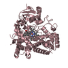
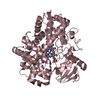
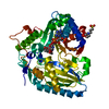
- PDB-4y8w: Crystal Structure of Human Cytochrome P450 21A2 Progesterone Complex -
+ Open data

- Basic information
| Entry | Database: PDB / ID: 4y8w | ||||||
|---|---|---|---|---|---|---|---|
| Title | Crystal Structure of Human Cytochrome P450 21A2 Progesterone Complex | ||||||
 Components Components | Cytochrome P450 21-hydroxylase | ||||||
 Keywords Keywords | OXIDOREDUCTASE / steroid hydroxylation / monooxygenases / adrenal steroidogenesis / congenital adrenal hyperplasia / Addison's disease / kinetic isotope effects | ||||||
| Function / homology |  Function and homology information Function and homology informationDefective CYP21A2 causes AH3 / steroid 21-monooxygenase / steroid 21-monooxygenase activity / 17-hydroxyprogesterone 21-hydroxylase activity / progesterone 21-hydroxylase activity / mineralocorticoid biosynthetic process / Mineralocorticoid biosynthesis / cortisol biosynthetic process / glucocorticoid biosynthetic process / Glucocorticoid biosynthesis ...Defective CYP21A2 causes AH3 / steroid 21-monooxygenase / steroid 21-monooxygenase activity / 17-hydroxyprogesterone 21-hydroxylase activity / progesterone 21-hydroxylase activity / mineralocorticoid biosynthetic process / Mineralocorticoid biosynthesis / cortisol biosynthetic process / glucocorticoid biosynthetic process / Glucocorticoid biosynthesis / sterol metabolic process / steroid biosynthetic process / steroid hydroxylase activity / steroid metabolic process / Endogenous sterols / steroid binding / iron ion binding / heme binding / endoplasmic reticulum membrane Similarity search - Function | ||||||
| Biological species |  Homo sapiens (human) Homo sapiens (human) | ||||||
| Method |  X-RAY DIFFRACTION / SYNCHROTRON / MOLECULAR REPLACEMENT / Resolution: 2.64 Å X-RAY DIFFRACTION / SYNCHROTRON / MOLECULAR REPLACEMENT / Resolution: 2.64 Å | ||||||
 Authors Authors | Pallan, P.S. / Lei, L. / Egli, M. | ||||||
| Funding support |  United States, 1items United States, 1items
| ||||||
 Citation Citation | Journal: J.Biol.Chem. / Year: 2015 Title: Human Cytochrome P450 21A2, the Major Steroid 21-Hydroxylase: STRUCTURE OF THE ENZYMEPROGESTERONE SUBSTRATE COMPLEX AND RATE-LIMITING C-H BOND CLEAVAGE. Authors: Pallan, P.S. / Wang, C. / Lei, L. / Yoshimoto, F.K. / Auchus, R.J. / Waterman, M.R. / Guengerich, F.P. / Egli, M. | ||||||
| History |
|
- Structure visualization
| Structure viewer | Molecule: MolmilJmol/JSmol |
|---|
- Downloads & links
-Download
| PDBx/mmCIF format | 4y8w.cif.gz | 276.6 KB | Display | PDBx/mmCIF format |
|---|---|---|---|---|
| PDB format | pdb4y8w.ent.gz | 221.3 KB | Display | PDB format |
| PDBx/mmJSON format | 4y8w.json.gz | Tree view | PDBx/mmJSON format | |
| Others |  Other downloads Other downloads |
-Validation report
| Arichive directory | https://data.pdbj.org/pub/pdb/validation_reports/y8/4y8wftp://data.pdbj.org/pub/pdb/validation_reports/y8/4y8w | HTTPS FTP |
|---|
-Related structure data
| Related structure data |  3qz1S S: Starting model for refinement |
|---|---|
| Similar structure data |


-Links
 PDBj
PDBj

- Assembly
| Deposited unit | 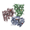
| ||||||||
|---|---|---|---|---|---|---|---|---|---|
| 1 | 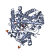
| ||||||||
| 2 | 
| ||||||||
| 3 | 
| ||||||||
| Unit cell |
|
-Components
| #1: Protein | Mass: 54539.160 Da / Num. of mol.: 3 / Fragment: residues 30-495 Source method: isolated from a genetically manipulated source Source: (gene. exp.) Homo sapiens (human) / Tissue: Adrenal cortex / Gene: P450-CYP21B, CYP21A2, hCG_1999926 / Organ: Kidneys / Plasmid: pET17b / Cell (production host): BL21-Gold DE(3) / Production host:  #2: Chemical | ChemComp-SO4 /   Mass: 96.063 Da / Num. of mol.: 4 / Source method: obtained synthetically / Formula: SO4 Mass: 96.063 Da / Num. of mol.: 4 / Source method: obtained synthetically / Formula: SO4#3: Chemical |   Mass: 616.487 Da / Num. of mol.: 3 Mass: 616.487 Da / Num. of mol.: 3Source method: isolated from a genetically manipulated source Formula: C34H32FeN4O4 #4: Chemical | 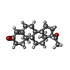  Mass: 314.462 Da / Num. of mol.: 3 Mass: 314.462 Da / Num. of mol.: 3Source method: isolated from a genetically manipulated source Formula: C21H30O2 #5: Water | ChemComp-HOH / |  Mass: 18.015 Da / Num. of mol.: 77 / Source method: isolated from a natural source / Formula: H2O Mass: 18.015 Da / Num. of mol.: 77 / Source method: isolated from a natural source / Formula: H2O |
|---|
-Experimental details
-Experiment
| Experiment | Method: X-RAY DIFFRACTION |
|---|
- Sample preparation
| Crystal | Density Matthews: 2.13 Å3/Da / Density % sol: 42.13 % |
|---|---|
| Crystal grow | Temperature: 291 K / Method: vapor diffusion, sitting drop / pH: 7.5 Details: 12 mg/mL protein, 0.05 M 4-(2-hydroxyethyl)-1-piperazineethanesulfonate (HEPES), 0.1 M ammonium sulfate and 12.5% (w/v) polyethylene glycol 3350 |
-Data collection
| Diffraction | Mean temperature: 100 K |
|---|---|
| Diffraction source | Source: SYNCHROTRON / Site: APS / Beamline: 21-ID-G / Wavelength: 0.9782 Å |
| Detector | Type: MARMOSAIC 300 mm CCD / Detector: CCD / Date: Aug 16, 2014 |
| Radiation | Protocol: SINGLE WAVELENGTH / Monochromatic (M) / Laue (L): M / Scattering type: x-ray |
| Radiation wavelength | Wavelength: 0.9782 Å / Relative weight: 1 |
| Reflection | Resolution: 2.64→30 Å / Num. obs: 40299 / % possible obs: 100 % / Observed criterion σ(I): 5 / Redundancy: 7.5 % / Net I/σ(I): 10.2 |
| Reflection shell | Resolution: 2.64→3.42 Å / Redundancy: 7.1 % / Mean I/σ(I) obs: 1.4 / % possible all: 100 |
- Processing
| Software |
| ||||||||||||||||||||||||||||||||||||||||||||||||||||||||||||||||||||||||||||||||||||||||||||||||||||||||||||||||||||||||||||||||||||||||||||||||||||||||||||||||||||||||||||||||||||||
|---|---|---|---|---|---|---|---|---|---|---|---|---|---|---|---|---|---|---|---|---|---|---|---|---|---|---|---|---|---|---|---|---|---|---|---|---|---|---|---|---|---|---|---|---|---|---|---|---|---|---|---|---|---|---|---|---|---|---|---|---|---|---|---|---|---|---|---|---|---|---|---|---|---|---|---|---|---|---|---|---|---|---|---|---|---|---|---|---|---|---|---|---|---|---|---|---|---|---|---|---|---|---|---|---|---|---|---|---|---|---|---|---|---|---|---|---|---|---|---|---|---|---|---|---|---|---|---|---|---|---|---|---|---|---|---|---|---|---|---|---|---|---|---|---|---|---|---|---|---|---|---|---|---|---|---|---|---|---|---|---|---|---|---|---|---|---|---|---|---|---|---|---|---|---|---|---|---|---|---|---|---|---|---|
| Refinement | Method to determine structure: MOLECULAR REPLACEMENT Starting model: PDB ID: 3QZ1 (One molecule of the protein alone) Resolution: 2.64→30 Å / Cor.coef. Fo:Fc: 0.916 / Cor.coef. Fo:Fc free: 0.874 / SU B: 18.477 / SU ML: 0.372 / Cross valid method: THROUGHOUT / ESU R Free: 0.411 / Stereochemistry target values: MAXIMUM LIKELIHOOD / Details: HYDROGENS HAVE BEEN USED IF PRESENT IN THE INPUT
| ||||||||||||||||||||||||||||||||||||||||||||||||||||||||||||||||||||||||||||||||||||||||||||||||||||||||||||||||||||||||||||||||||||||||||||||||||||||||||||||||||||||||||||||||||||||
| Solvent computation | Ion probe radii: 0.8 Å / Shrinkage radii: 0.8 Å / VDW probe radii: 1.2 Å / Solvent model: MASK | ||||||||||||||||||||||||||||||||||||||||||||||||||||||||||||||||||||||||||||||||||||||||||||||||||||||||||||||||||||||||||||||||||||||||||||||||||||||||||||||||||||||||||||||||||||||
| Displacement parameters | Biso mean: 42.618 Å2
| ||||||||||||||||||||||||||||||||||||||||||||||||||||||||||||||||||||||||||||||||||||||||||||||||||||||||||||||||||||||||||||||||||||||||||||||||||||||||||||||||||||||||||||||||||||||
| Refinement step | Cycle: LAST / Resolution: 2.64→30 Å
| ||||||||||||||||||||||||||||||||||||||||||||||||||||||||||||||||||||||||||||||||||||||||||||||||||||||||||||||||||||||||||||||||||||||||||||||||||||||||||||||||||||||||||||||||||||||
| Refine LS restraints |
|