 Movie
Movie Controller

[English] 日本語
 Yorodumi
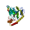
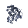
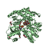
- PDB-3hgk: crystal structure of effect protein AvrptoB complexed with kinase Pto -
+ Open data

- Basic information
| Entry | Database: PDB / ID: 3hgk | ||||||
|---|---|---|---|---|---|---|---|
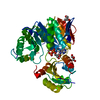
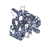
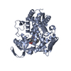
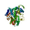
| Title | crystal structure of effect protein AvrptoB complexed with kinase Pto | ||||||
 Components Components |
| ||||||
 Keywords Keywords | TRANSFERASE / five helices / Pto P+1 loop / ATP-binding / Kinase / Nucleotide-binding / Serine/threonine-protein kinase / Hypersensitive response elicitation / Ligase / Secreted / Ubl conjugation / Ubl conjugation pathway / Virulence | ||||||
| Function / homology |  Function and homology information Function and homology informationeffector-mediated suppression of host pattern-triggered immunity / symbiont-mediated perturbation of host programmed cell death / plasmodesma / Transferases; Acyltransferases; Aminoacyltransferases / transmembrane receptor protein tyrosine kinase activity / transferase activity / protein serine/threonine kinase activity / extracellular region / ATP binding / plasma membrane Similarity search - Function | ||||||
| Biological species |   Pseudomonas syringae pv. tomato (bacteria) Pseudomonas syringae pv. tomato (bacteria) | ||||||
| Method |  X-RAY DIFFRACTION / SYNCHROTRON / MOLECULAR REPLACEMENT / Resolution: 3.3 Å X-RAY DIFFRACTION / SYNCHROTRON / MOLECULAR REPLACEMENT / Resolution: 3.3 Å | ||||||
 Authors Authors | Dong, J. / Fan, F. / Gu, L. / Chai, J. | ||||||
 Citation Citation | Journal: Plant Cell / Year: 2009 Title: Crystal Structure of the Complex between Pseudomonas Effector AvrPtoB and the Tomato Pto Kinase Reveals Both a Shared and a Unique Interface Compared with AvrPto-Pto Authors: Dong, J. / Xiao, F. / Fan, F. / Gu, L. / Cang, H. / Martin, G.B. / Chai, J. | ||||||
| History |
|
- Structure visualization
| Structure viewer | Molecule: MolmilJmol/JSmol |
|---|
- Downloads & links
-Download
| PDBx/mmCIF format | 3hgk.cif.gz | 291.7 KB | Display | PDBx/mmCIF format |
|---|---|---|---|---|
| PDB format | pdb3hgk.ent.gz | 238.1 KB | Display | PDB format |
| PDBx/mmJSON format | 3hgk.json.gz | Tree view | PDBx/mmJSON format | |
| Others |  Other downloads Other downloads |
-Validation report
| Arichive directory | https://data.pdbj.org/pub/pdb/validation_reports/hg/3hgkftp://data.pdbj.org/pub/pdb/validation_reports/hg/3hgk | HTTPS FTP |
|---|
-Related structure data
| Related structure data | 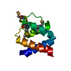 3hglSC  2qkwS S: Starting model for refinement C: citing same article ( |
|---|---|
| Similar structure data |


-Links
 PDBj
PDBj- Assembly
| Deposited unit | 
| ||||||||
|---|---|---|---|---|---|---|---|---|---|
| 1 | 
| ||||||||
| 2 | 
| ||||||||
| 3 | 
| ||||||||
| 4 | 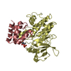
| ||||||||
| Unit cell |
|
-Components
| #1: Protein | Mass: 37330.395 Da / Num. of mol.: 4 / Mutation: D193G Source method: isolated from a genetically manipulated source Source: (gene. exp.) Gene: Pto / Plasmid: PET-30A / Production host: #2: Protein | Mass: 9428.645 Da / Num. of mol.: 4 / Fragment: UNP residues 121-205 Source method: isolated from a genetically manipulated source Source: (gene. exp.) Pseudomonas syringae pv. tomato (bacteria)Gene: hopAB2, avrPtoB, PSPTO_3087 / Species (production host): plasmid / Production host: References: UniProt: Q8RSY1, Ligases; Forming carbon-nitrogen bonds; Acid-amino-acid ligases (peptide synthases) Has protein modification | Y | |
|---|
-Experimental details
-Experiment
| Experiment | Method: X-RAY DIFFRACTION / Number of used crystals: 2 |
|---|
- Sample preparation
| Crystal | Density Matthews: 2.55 Å3/Da / Density % sol: 51.74 % |
|---|---|
| Crystal grow | Temperature: 293 K / Method: vapor diffusion, hanging drop / pH: 7.9 Details: 0.1M tri-Sodium Citrate dihydrate, 17.5% (w/v) Polyethylene Glycol 3350, 0.1mM Tris-HCl pH 7.9, 10.0mM phenol, VAPOR DIFFUSION, HANGING DROP, temperature 293K |
-Data collection
| Diffraction | Mean temperature: 100 K |
|---|---|
| Diffraction source | Source: SYNCHROTRON / Site: Photon Factory  / Beamline: AR-NW12A / Wavelength: 1 Å / Beamline: AR-NW12A / Wavelength: 1 Å |
| Detector | Type: MAR CCD 165 mm / Detector: CCD / Date: Jan 7, 2008 |
| Radiation | Monochromator: Si(111) double-crystal / Protocol: SINGLE WAVELENGTH / Monochromatic (M) / Laue (L): M / Scattering type: x-ray |
| Radiation wavelength | Wavelength: 1 Å / Relative weight: 1 |
| Reflection | Resolution: 3.3→99 Å / Num. obs: 29886 / % possible obs: 99.8 % / Observed criterion σ(F): 2 / Observed criterion σ(I): 2.9 / Redundancy: 5.6 % / Rmerge(I) obs: 0.081 / Net I/σ(I): 22.3 |
| Reflection shell | Resolution: 3.3→3.37 Å / Redundancy: 5.3 % / Rmerge(I) obs: 0.53 / Mean I/σ(I) obs: 2.9 / % possible all: 99.6 |
- Processing
| Software |
| ||||||||||||||||||||||||||||||||||||||||||||||||||||||||||||||||||||||||||||||||||||||||||
|---|---|---|---|---|---|---|---|---|---|---|---|---|---|---|---|---|---|---|---|---|---|---|---|---|---|---|---|---|---|---|---|---|---|---|---|---|---|---|---|---|---|---|---|---|---|---|---|---|---|---|---|---|---|---|---|---|---|---|---|---|---|---|---|---|---|---|---|---|---|---|---|---|---|---|---|---|---|---|---|---|---|---|---|---|---|---|---|---|---|---|---|
| Refinement | Method to determine structure: MOLECULAR REPLACEMENT Starting model: PDB ENTRIES 3HGL for AvrPtoB AND 2QKW for Pto Resolution: 3.3→20 Å / Cor.coef. Fo:Fc: 0.916 / Cor.coef. Fo:Fc free: 0.907 / Occupancy max: 1 / Occupancy min: 1 / SU B: 120.055 / SU ML: 0.79 / Cross valid method: THROUGHOUT / σ(F): 0 / ESU R Free: 0.714 / Stereochemistry target values: MAXIMUM LIKELIHOOD
| ||||||||||||||||||||||||||||||||||||||||||||||||||||||||||||||||||||||||||||||||||||||||||
| Solvent computation | Ion probe radii: 0.8 Å / Shrinkage radii: 0.8 Å / VDW probe radii: 1.1 Å / Solvent model: MASK | ||||||||||||||||||||||||||||||||||||||||||||||||||||||||||||||||||||||||||||||||||||||||||
| Displacement parameters | Biso max: 119.92 Å2 / Biso mean: 81.806 Å2 / Biso min: 44.12 Å2
| ||||||||||||||||||||||||||||||||||||||||||||||||||||||||||||||||||||||||||||||||||||||||||
| Refinement step | Cycle: LAST / Resolution: 3.3→20 Å
| ||||||||||||||||||||||||||||||||||||||||||||||||||||||||||||||||||||||||||||||||||||||||||
| Refine LS restraints |
| ||||||||||||||||||||||||||||||||||||||||||||||||||||||||||||||||||||||||||||||||||||||||||
| LS refinement shell | Resolution: 3.297→3.38 Å / Total num. of bins used: 20
|
