 Movie
Movie Controller

[English] 日本語
 Yorodumi
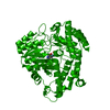
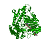
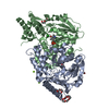
- PDB-1gey: CRYSTAL STRUCTURE OF HISTIDINOL-PHOSPHATE AMINOTRANSFERASE COMPLE... -
+ Open data

- Basic information
| Entry | Database: PDB / ID: 1gey | ||||||
|---|---|---|---|---|---|---|---|
| Title | CRYSTAL STRUCTURE OF HISTIDINOL-PHOSPHATE AMINOTRANSFERASE COMPLEXED WITH N-(5'-PHOSPHOPYRIDOXYL)-L-GLUTAMATE | ||||||
 Components Components | HISTIDINOL-PHOSPHATE AMINOTRANSFERASE | ||||||
 Keywords Keywords | TRANSFERASE / alpha/beta-structure / N-(5'-PHOSPHOPYRIDOXYL)-L-GLUTAMATE / PPE / complex | ||||||
| Function / homology |  Function and homology information Function and homology informationhistidinol-phosphate transaminase / L-histidinol-phosphate:2-oxoglutarate transaminase activity / L-histidine biosynthetic process / pyridoxal phosphate binding / identical protein binding / cytosol Similarity search - Function | ||||||
| Biological species |  | ||||||
| Method |  X-RAY DIFFRACTION / FOURIER SYNTHESIS / Resolution: 2.3 Å X-RAY DIFFRACTION / FOURIER SYNTHESIS / Resolution: 2.3 Å | ||||||
 Authors Authors | Haruyama, K. / Nakai, T. / Miyahara, I. / Hirotsu, K. / Mizuguchi, H. / Hayashi, H. / Kagamiyama, H. | ||||||
 Citation Citation | Journal: Biochemistry / Year: 2001 Title: Structures of Escherichia coli histidinol-phosphate aminotransferase and its complexes with histidinol-phosphate and N-(5'-phosphopyridoxyl)-L-glutamate: double substrate recognition of the enzyme. Authors: Haruyama, K. / Nakai, T. / Miyahara, I. / Hirotsu, K. / Mizuguchi, H. / Hayashi, H. / Kagamiyama, H. | ||||||
| History |
|
- Structure visualization
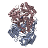
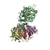
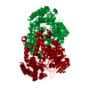
| Structure viewer | Molecule: MolmilJmol/JSmol |
|---|
- Downloads & links
-Download
| PDBx/mmCIF format | 1gey.cif.gz | 81.6 KB | Display | PDBx/mmCIF format |
|---|---|---|---|---|
| PDB format | pdb1gey.ent.gz | 59.8 KB | Display | PDB format |
| PDBx/mmJSON format | 1gey.json.gz | Tree view | PDBx/mmJSON format | |
| Others |  Other downloads Other downloads |
-Validation report
| Arichive directory | https://data.pdbj.org/pub/pdb/validation_reports/ge/1geyftp://data.pdbj.org/pub/pdb/validation_reports/ge/1gey | HTTPS FTP |
|---|
-Related structure data
| Related structure data |  1gewSC  1gexC S: Starting model for refinement C: citing same article ( |
|---|---|
| Similar structure data |


-Links
 PDBj
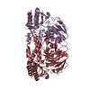
PDBj- Assembly
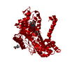
| Deposited unit | 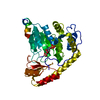
| ||||||||
|---|---|---|---|---|---|---|---|---|---|
| 1 | 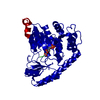
| ||||||||
| Unit cell |
| ||||||||
| Details | The biological assembly is a dimer constructed from chain A a symmetry partner generated by the two-fold. |
-Components
| #1: Protein | Mass: 39393.980 Da / Num. of mol.: 1 Source method: isolated from a genetically manipulated source Source: (gene. exp.) References: UniProt: P06986, histidinol-phosphate transaminase |
|---|---|
| #2: Chemical | ChemComp-PPE /   Mass: 379.280 Da / Num. of mol.: 1 / Source method: obtained synthetically / Formula: C13H20N2O9P Mass: 379.280 Da / Num. of mol.: 1 / Source method: obtained synthetically / Formula: C13H20N2O9P |
| #3: Water | ChemComp-HOH /  Mass: 18.015 Da / Num. of mol.: 116 / Source method: isolated from a natural source / Formula: H2O Mass: 18.015 Da / Num. of mol.: 116 / Source method: isolated from a natural source / Formula: H2O |
-Experimental details
-Experiment
| Experiment | Method: X-RAY DIFFRACTION / Number of used crystals: 1 |
|---|
- Sample preparation
| Crystal | Density Matthews: 2.41 Å3/Da / Density % sol: 48.99 % | ||||||||||||||||||||||||||||||
|---|---|---|---|---|---|---|---|---|---|---|---|---|---|---|---|---|---|---|---|---|---|---|---|---|---|---|---|---|---|---|---|
| Crystal grow | Temperature: 293 K / Method: vapor diffusion, hanging drop / pH: 7.7 Details: PEG 4000, magnesium chloride, tris, pH 7.7, VAPOR DIFFUSION, HANGING DROP, temperature 293.0K | ||||||||||||||||||||||||||||||
| Crystal grow | *PLUS Temperature: 20 ℃ | ||||||||||||||||||||||||||||||
| Components of the solutions | *PLUS
|
-Data collection
| Diffraction | Mean temperature: 287 K |
|---|---|
| Diffraction source | Source: ROTATING ANODE / Type: RIGAKU RU200 / Wavelength: 1.5418 |
| Detector | Type: RIGAKU RAXIS IIC / Detector: IMAGE PLATE / Date: Sep 26, 2000 |
| Radiation | Protocol: SINGLE WAVELENGTH / Monochromatic (M) / Laue (L): M / Scattering type: x-ray |
| Radiation wavelength | Wavelength: 1.5418 Å / Relative weight: 1 |
| Reflection | Resolution: 2.3→50 Å / Num. all: 55982 / Num. obs: 55982 / % possible obs: 99.1 % / Observed criterion σ(F): 0 / Observed criterion σ(I): 0 / Redundancy: 3.4 % / Biso Wilson estimate: 30.225 Å2 / Rmerge(I) obs: 0.089 / Net I/σ(I): 10.17 |
| Reflection shell | Resolution: 2.3→2.38 Å / Redundancy: 2.8 % / Rmerge(I) obs: 0.265 / Num. unique all: 1628 / % possible all: 97.4 |
| Reflection | *PLUS Num. obs: 16231 / Num. measured all: 55982 |
| Reflection shell | *PLUS % possible obs: 97.4 % |
- Processing
| Software |
| |||||||||||||||||||||||||
|---|---|---|---|---|---|---|---|---|---|---|---|---|---|---|---|---|---|---|---|---|---|---|---|---|---|---|
| Refinement | Method to determine structure: FOURIER SYNTHESIS Starting model: 1GEW Resolution: 2.3→8 Å / σ(F): 2 / σ(I): 0 / Stereochemistry target values: Engh & Huber
| |||||||||||||||||||||||||
| Refinement step | Cycle: LAST / Resolution: 2.3→8 Å
| |||||||||||||||||||||||||
| Refine LS restraints |
| |||||||||||||||||||||||||
| Software | *PLUS Name: X-PLOR / Version: 3.851 / Classification: refinement | |||||||||||||||||||||||||
| LS refinement shell | *PLUS Rfactor Rfree: 0.349 / Rfactor Rwork: 0.24 |
