 Movie
Movie Controller

[English] 日本語
 Yorodumi
- PDB-1yjs: K226Q Mutant Of Serine Hydroxymethyltransferase From B. Stearothe... -
+ Open data

- Basic information
| Entry | Database: PDB / ID: 1yjs | ||||||
|---|---|---|---|---|---|---|---|
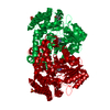
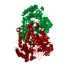
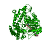
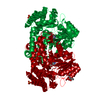
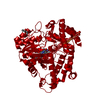
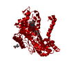
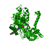
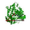
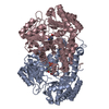
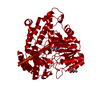
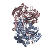
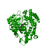
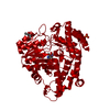
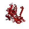
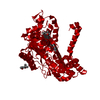
| Title | K226Q Mutant Of Serine Hydroxymethyltransferase From B. Stearothermophilus, Complex With Glycine | ||||||
 Components Components | SERINE HYDROXYMETHYLTRANSFERASE | ||||||
 Keywords Keywords | TRANSFERASE / SHMT / Mutant / Catalysis | ||||||
| Function / homology |  Function and homology information Function and homology informationglycine hydroxymethyltransferase / glycine hydroxymethyltransferase activity / : / tetrahydrofolate interconversion / pyridoxal phosphate binding / cytosol Similarity search - Function | ||||||
| Biological species |   Geobacillus stearothermophilus (bacteria) Geobacillus stearothermophilus (bacteria) | ||||||
| Method |  X-RAY DIFFRACTION / MOLECULAR REPLACEMENT / Resolution: 2 Å X-RAY DIFFRACTION / MOLECULAR REPLACEMENT / Resolution: 2 Å | ||||||
 Authors Authors | Bhavani, S. / Trivedi, V. / Jala, V.R. / Subramanya, H.S. / Kaul, P. / Purnima, K. / Prakash, V. / Appaji, R.N. / Savithri, H.S. | ||||||
 Citation Citation | Journal: Biochemistry / Year: 2005 Title: Role of Lys-226 in the Catalytic Mechanism of Bacillus Stearothermophilus Serine Hydroxymethyltransferase-Crystal Structure and Kinetic Studies Authors: Bhavani, S. / Trivedi, V. / Jala, V.R. / Subramanya, H.S. / Kaul, P. / Purnima, K. / Prakash, V. / Appaji, R.N. / Savithri, H.S. | ||||||
| History |
|
- Structure visualization
| Structure viewer | Molecule: MolmilJmol/JSmol |
|---|
- Downloads & links
-Download
| PDBx/mmCIF format | 1yjs.cif.gz | 92.9 KB | Display | PDBx/mmCIF format |
|---|---|---|---|---|
| PDB format | pdb1yjs.ent.gz | 69.9 KB | Display | PDB format |
| PDBx/mmJSON format | 1yjs.json.gz | Tree view | PDBx/mmJSON format | |
| Others |  Other downloads Other downloads |
-Validation report
| Arichive directory | https://data.pdbj.org/pub/pdb/validation_reports/yj/1yjsftp://data.pdbj.org/pub/pdb/validation_reports/yj/1yjs | HTTPS FTP |
|---|
-Related structure data
| Related structure data |  1yjyC  1yjzC 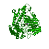 1kkjS C: citing same article ( S: Starting model for refinement |
|---|---|
| Similar structure data |


-Links
 PDBj
PDBj

- Assembly
| Deposited unit | 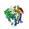
| ||||||||
|---|---|---|---|---|---|---|---|---|---|
| 1 | 
| ||||||||
| Unit cell |
| ||||||||
| Details | The second part of the biological assembly is generated by the two fold axis : -X, -Y, Z. |
-Components
| #1: Protein | Mass: 45710.688 Da / Num. of mol.: 1 / Fragment: Serine methylase / Mutation: K226Q Source method: isolated from a genetically manipulated source Source: (gene. exp.) Geobacillus stearothermophilus (bacteria)Gene: SHMT / Plasmid: PRSETC / Species (production host): Escherichia coli / Production host: References: UniProt: Q7SIB6, glycine hydroxymethyltransferase |
|---|---|
| #2: Chemical | ChemComp-PLP /   Mass: 247.142 Da / Num. of mol.: 1 / Source method: obtained synthetically / Formula: C8H10NO6P Mass: 247.142 Da / Num. of mol.: 1 / Source method: obtained synthetically / Formula: C8H10NO6P |
| #3: Chemical | ChemComp-GLY /   Type: peptide linking / Mass: 75.067 Da / Num. of mol.: 1 / Source method: obtained synthetically / Formula: C2H5NO2 Type: peptide linking / Mass: 75.067 Da / Num. of mol.: 1 / Source method: obtained synthetically / Formula: C2H5NO2 |
| #4: Water | ChemComp-HOH /  Mass: 18.015 Da / Num. of mol.: 139 / Source method: isolated from a natural source / Formula: H2O Mass: 18.015 Da / Num. of mol.: 139 / Source method: isolated from a natural source / Formula: H2O |
-Experimental details
-Experiment
| Experiment | Method: X-RAY DIFFRACTION / Number of used crystals: 1 |
|---|
- Sample preparation
| Crystal | Density Matthews: 2.03 Å3/Da / Density % sol: 39.49 % |
|---|---|
| Crystal grow | Temperature: 298 K / Method: vapor diffusion, hanging drop / pH: 7.5 Details: Hepes, MPD, pH 7.5, VAPOR DIFFUSION, HANGING DROP, temperature 298.0K |
-Data collection
| Diffraction | Mean temperature: 100 K |
|---|---|
| Diffraction source | Source: ROTATING ANODE / Type: RIGAKU RU300 / Wavelength: 1.5418 Å |
| Detector | Type: MARRESEARCH / Detector: IMAGE PLATE / Date: Mar 11, 2002 / Details: Mirrors |
| Radiation | Monochromator: Ni Filter / Protocol: SINGLE WAVELENGTH / Monochromatic (M) / Laue (L): M / Scattering type: x-ray |
| Radiation wavelength | Wavelength: 1.5418 Å / Relative weight: 1 |
| Reflection | Resolution: 2→15 Å / Num. all: 25927 / Num. obs: 25927 / % possible obs: 97.5 % / Observed criterion σ(F): -3 / Observed criterion σ(I): -3 / Redundancy: 2.4 % / Net I/σ(I): 20.7 |
| Reflection shell | Resolution: 2→2.07 Å / Redundancy: 1.6 % / Rmerge(I) obs: 0.149 / Num. unique all: 2481 / % possible all: 98.7 |
- Processing
| Software |
| |||||||||||||||
|---|---|---|---|---|---|---|---|---|---|---|---|---|---|---|---|---|
| Refinement | Method to determine structure: MOLECULAR REPLACEMENT Starting model: 1KKJ Resolution: 2→10 Å / Isotropic thermal model: Anisoltrophic / σ(F): 0 / σ(I): 3 / Stereochemistry target values: Engh & Huber
| |||||||||||||||
| Refinement step | Cycle: LAST / Resolution: 2→10 Å
| |||||||||||||||
| Refine LS restraints |
| |||||||||||||||
| LS refinement shell | Resolution: 2→2.07 Å / Rfactor Rfree error: 0.22084
|
