 Movie
Movie Controller

[English] 日本語
 Yorodumi
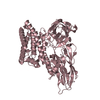
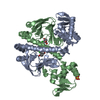
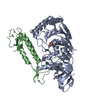
- PDB-1bp3: THE XRAY STRUCTURE OF A GROWTH HORMONE-PROLACTIN RECEPTOR COMPLEX -
+ Open data

- Basic information
| Entry | Database: PDB / ID: 1bp3 | ||||||
|---|---|---|---|---|---|---|---|
| Title | THE XRAY STRUCTURE OF A GROWTH HORMONE-PROLACTIN RECEPTOR COMPLEX | ||||||
 Components Components |
| ||||||
 Keywords Keywords | HORMONE/GROWTH FACTOR / HORMONE / RECEPTOR / HORMONE-GROWTH FACTOR complex | ||||||
| Function / homology |  Function and homology information Function and homology informationgrowth hormone activity / growth hormone receptor complex / prolactin receptor activity / prolactin receptor binding / bone maturation / activation of transmembrane receptor protein tyrosine kinase activity / animal organ development / positive regulation of multicellular organism growth / steroid biosynthetic process / activation of Janus kinase activity ...growth hormone activity / growth hormone receptor complex / prolactin receptor activity / prolactin receptor binding / bone maturation / activation of transmembrane receptor protein tyrosine kinase activity / animal organ development / positive regulation of multicellular organism growth / steroid biosynthetic process / activation of Janus kinase activity / cellular response to granulocyte macrophage colony-stimulating factor stimulus / positive regulation of insulin-like growth factor receptor signaling pathway / positive regulation of D-glucose transmembrane transport / growth hormone receptor binding / cell surface receptor signaling pathway via STAT / growth hormone receptor signaling pathway / Prolactin receptor signaling / cytokine binding / peptide hormone binding / growth hormone receptor signaling pathway via JAK-STAT / Synthesis, secretion, and deacylation of Ghrelin / Growth hormone receptor signaling / cell surface receptor signaling pathway via JAK-STAT / lactation / embryo implantation / positive regulation of B cell proliferation / cytokine activity / endosome lumen / positive regulation of receptor signaling pathway via JAK-STAT / growth factor activity / hormone activity / response to nutrient levels / cytokine-mediated signaling pathway / response to estradiol / positive regulation of cold-induced thermogenesis / positive regulation of MAPK cascade / positive regulation of phosphatidylinositol 3-kinase/protein kinase B signal transduction / signaling receptor complex / external side of plasma membrane / positive regulation of cell population proliferation / lipid binding / negative regulation of apoptotic process / protein kinase binding / cell surface / : / extracellular region / metal ion binding / plasma membrane Similarity search - Function | ||||||
| Biological species |  Homo sapiens (human) Homo sapiens (human) | ||||||
| Method |  X-RAY DIFFRACTION / SYNCHROTRON / MOLECULAR REPLACEMENT / Resolution: 2.9 Å X-RAY DIFFRACTION / SYNCHROTRON / MOLECULAR REPLACEMENT / Resolution: 2.9 Å | ||||||
 Authors Authors | Somers, W. / Ultsch, M. / De Vos, A.M. / Kossiakoff, A.A. | ||||||
 Citation Citation | Journal: Nature / Year: 1994 Title: The X-ray structure of a growth hormone-prolactin receptor complex. Authors: Somers, W. / Ultsch, M. / De Vos, A.M. / Kossiakoff, A.A. | ||||||
| History |
|
- Structure visualization
| Structure viewer | Molecule: MolmilJmol/JSmol |
|---|
- Downloads & links
-Download
| PDBx/mmCIF format | 1bp3.cif.gz | 91.2 KB | Display | PDBx/mmCIF format |
|---|---|---|---|---|
| PDB format | pdb1bp3.ent.gz | 68.2 KB | Display | PDB format |
| PDBx/mmJSON format | 1bp3.json.gz | Tree view | PDBx/mmJSON format | |
| Others |  Other downloads Other downloads |
-Validation report
| Arichive directory | https://data.pdbj.org/pub/pdb/validation_reports/bp/1bp3ftp://data.pdbj.org/pub/pdb/validation_reports/bp/1bp3 | HTTPS FTP |
|---|
-Related structure data
| Related structure data |  1a22S S: Starting model for refinement |
|---|---|
| Similar structure data |


-Links
 PDBj
PDBj


- Assembly
| Deposited unit | 
| ||||||||
|---|---|---|---|---|---|---|---|---|---|
| 1 |
| ||||||||
| Unit cell |
|
-Components
| #1: Protein | Mass: 22252.135 Da / Num. of mol.: 1 / Mutation: G120R Source method: isolated from a genetically manipulated source Source: (gene. exp.) Homo sapiens (human) / Production host:  |
|---|---|
| #2: Protein | Mass: 24543.875 Da / Num. of mol.: 1 Source method: isolated from a genetically manipulated source Source: (gene. exp.) Homo sapiens (human) / Production host: |
| #3: Chemical | ChemComp-ZN /   Mass: 65.409 Da / Num. of mol.: 1 / Source method: obtained synthetically / Formula: Zn Mass: 65.409 Da / Num. of mol.: 1 / Source method: obtained synthetically / Formula: Zn |
| Has protein modification | Y |
-Experimental details
-Experiment
| Experiment | Method: X-RAY DIFFRACTION / Number of used crystals: 9 |
|---|
- Sample preparation
| Crystal | Density Matthews: 2.49 Å3/Da / Density % sol: 50.57 % |
|---|---|
| Crystal grow | pH: 6 / Details: pH 6.00 |
-Data collection
| Diffraction | Mean temperature: 300 K |
|---|---|
| Diffraction source | Source: SYNCHROTRON / Site: SSRL  / Beamline: BL7-1 / Wavelength: 0.908 / Beamline: BL7-1 / Wavelength: 0.908 |
| Detector | Type: MARRESEARCH / Detector: IMAGE PLATE |
| Radiation | Protocol: SINGLE WAVELENGTH / Monochromatic (M) / Laue (L): M / Scattering type: x-ray |
| Radiation wavelength | Wavelength: 0.908 Å / Relative weight: 1 |
| Reflection | Resolution: 2.9→10 Å / Num. obs: 9883 / % possible obs: 93 % / Rmerge(I) obs: 0.108 |
| Reflection shell | Resolution: 2.9→3 Å / % possible all: 89 |
- Processing
| Software |
| |||||||||||||||||||||||||||||||||||||||||||||||||||||||||||||||
|---|---|---|---|---|---|---|---|---|---|---|---|---|---|---|---|---|---|---|---|---|---|---|---|---|---|---|---|---|---|---|---|---|---|---|---|---|---|---|---|---|---|---|---|---|---|---|---|---|---|---|---|---|---|---|---|---|---|---|---|---|---|---|---|---|
| Refinement | Method to determine structure: MOLECULAR REPLACEMENT Starting model: PDB ENTRY 1A22 Resolution: 2.9→10 Å / σ(F): 0
| |||||||||||||||||||||||||||||||||||||||||||||||||||||||||||||||
| Refinement step | Cycle: LAST / Resolution: 2.9→10 Å
| |||||||||||||||||||||||||||||||||||||||||||||||||||||||||||||||
| Refine LS restraints |
|
