 Movie
Movie Controller

[English] 日本語
 Yorodumi
- PDB-5oht: A GH31 family sulfoquinovosidase from E. coli in complex with aza... -
+ Open data

- Basic information
| Entry | Database: PDB / ID: 5oht | ||||||
|---|---|---|---|---|---|---|---|
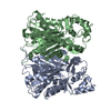
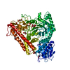
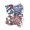
| Title | A GH31 family sulfoquinovosidase from E. coli in complex with aza-sugar inhibitor IFGSQ | ||||||
 Components Components | Sulfoquinovosidase | ||||||
 Keywords Keywords | HYDROLASE / sulfoglycosidase / sulfoglycolysis / complex / general acid-base varient | ||||||
| Function / homology |  Function and homology information Function and homology informationsulfoquinovosidase / sulfoquinovosidase activity / : / hydrolase activity, hydrolyzing O-glycosyl compounds / carbohydrate binding / carbohydrate metabolic process Similarity search - Function | ||||||
| Biological species |  | ||||||
| Method |  X-RAY DIFFRACTION / SYNCHROTRON / MOLECULAR REPLACEMENT / Resolution: 1.87 Å X-RAY DIFFRACTION / SYNCHROTRON / MOLECULAR REPLACEMENT / Resolution: 1.87 Å | ||||||
 Authors Authors | Jin, Y. / Williams, S.J. / Goddard-Borger, E. / Davies, G.J. | ||||||
 Citation Citation | Journal: ACS Cent Sci / Year: 2018 Title: Structural and Biochemical Insights into the Function and Evolution of Sulfoquinovosidases. Authors: Abayakoon, P. / Jin, Y. / Lingford, J.P. / Petricevic, M. / John, A. / Ryan, E. / Wai-Ying Mui, J. / Pires, D.E.V. / Ascher, D.B. / Davies, G.J. / Goddard-Borger, E.D. / Williams, S.J. | ||||||
| History |
|
- Structure visualization
| Structure viewer | Molecule: MolmilJmol/JSmol |
|---|
- Downloads & links
-Download
| PDBx/mmCIF format | 5oht.cif.gz | 291.3 KB | Display | PDBx/mmCIF format |
|---|---|---|---|---|
| PDB format | pdb5oht.ent.gz | 231.8 KB | Display | PDB format |
| PDBx/mmJSON format | 5oht.json.gz | Tree view | PDBx/mmJSON format | |
| Others |  Other downloads Other downloads |
-Validation report
| Arichive directory | https://data.pdbj.org/pub/pdb/validation_reports/oh/5ohtftp://data.pdbj.org/pub/pdb/validation_reports/oh/5oht | HTTPS FTP |
|---|
-Related structure data
| Related structure data | 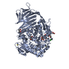 5ohsC 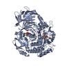 5ohyC 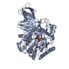 5aeeS S: Starting model for refinement C: citing same article ( |
|---|---|
| Similar structure data |


-Links
 PDBj
PDBj


- Assembly
| Deposited unit | 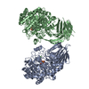
| ||||||||
|---|---|---|---|---|---|---|---|---|---|
| 1 | 
| ||||||||
| 2 | 
| ||||||||
| Unit cell |
| ||||||||
| Details | biological unit is the same as asym. |
-Components
| #1: Protein | Mass: 78423.898 Da / Num. of mol.: 2 Source method: isolated from a genetically manipulated source Source: (gene. exp.) Gene: yihQ, squQ, b3878, JW3849 / Production host: #2: Chemical |   Mass: 40.078 Da / Num. of mol.: 2 / Source method: obtained synthetically / Formula: Ca Mass: 40.078 Da / Num. of mol.: 2 / Source method: obtained synthetically / Formula: Ca#3: Chemical |   Mass: 211.236 Da / Num. of mol.: 2 / Source method: obtained synthetically / Formula: C6H13NO5S Mass: 211.236 Da / Num. of mol.: 2 / Source method: obtained synthetically / Formula: C6H13NO5S#4: Water | ChemComp-HOH / |  Mass: 18.015 Da / Num. of mol.: 618 / Source method: isolated from a natural source / Formula: H2O Mass: 18.015 Da / Num. of mol.: 618 / Source method: isolated from a natural source / Formula: H2O |
|---|
-Experimental details
-Experiment
| Experiment | Method: X-RAY DIFFRACTION / Number of used crystals: 1 |
|---|
- Sample preparation
| Crystal | Density Matthews: 2.9 Å3/Da / Density % sol: 57.9 % |
|---|---|
| Crystal grow | Temperature: 293 K / Method: vapor diffusion, sitting drop Details: 25 MG/ML-1 PROTEIN IN 50 MM NAPO4 , PH6.5, AND 250 MM NACL BUFFER IS MIXED WITH EQUAL VOLUME OF PRECIPITANT COMPOSED OF 50-60% (V/V) 2-METHYL-2, 4-PENTANEDIOL, 0.1-0.15 M CACL2, AND 20 DEGREE |
-Data collection
| Diffraction | Mean temperature: 100 K |
|---|---|
| Diffraction source | Source: SYNCHROTRON / Site: Diamond  / Beamline: I04-1 / Wavelength: 0.92819 Å / Beamline: I04-1 / Wavelength: 0.92819 Å |
| Detector | Type: DECTRIS PILATUS 6M-F / Detector: PIXEL / Date: Dec 12, 2016 |
| Radiation | Protocol: SINGLE WAVELENGTH / Monochromatic (M) / Laue (L): M / Scattering type: x-ray |
| Radiation wavelength | Wavelength: 0.92819 Å / Relative weight: 1 |
| Reflection | Resolution: 1.87→44.62 Å / Num. obs: 140829 / % possible obs: 97.8 % / Redundancy: 3.5 % / CC1/2: 0.999 / Rmerge(I) obs: 0.0378 / Net I/σ(I): 14.8 |
| Reflection shell | Resolution: 1.87→1.9 Å / Redundancy: 3.6 % / Rmerge(I) obs: 0.797 / Mean I/σ(I) obs: 1.2 / Num. unique obs: 6956 / CC1/2: 0.622 / % possible all: 96.9 |
- Processing
| Software |
| |||||||||||||||||||||||||||||||||||||||||||||||||||||||||||||||||||||||||||
|---|---|---|---|---|---|---|---|---|---|---|---|---|---|---|---|---|---|---|---|---|---|---|---|---|---|---|---|---|---|---|---|---|---|---|---|---|---|---|---|---|---|---|---|---|---|---|---|---|---|---|---|---|---|---|---|---|---|---|---|---|---|---|---|---|---|---|---|---|---|---|---|---|---|---|---|---|
| Refinement | Method to determine structure: MOLECULAR REPLACEMENT Starting model: 5aee Resolution: 1.87→44.62 Å / Cor.coef. Fo:Fc: 0.975 / Cor.coef. Fo:Fc free: 0.964 / Cross valid method: THROUGHOUT / σ(F): 0 / ESU R: 0.114 / ESU R Free: 0.113 Details: HYDROGENS HAVE BEEN ADDED IN THE RIDING POSITIONS U VALUES : REFINED INDIVIDUALLY
| |||||||||||||||||||||||||||||||||||||||||||||||||||||||||||||||||||||||||||
| Solvent computation | Ion probe radii: 0.8 Å / Shrinkage radii: 0.8 Å / VDW probe radii: 1.2 Å | |||||||||||||||||||||||||||||||||||||||||||||||||||||||||||||||||||||||||||
| Displacement parameters | Biso max: 117.14 Å2 / Biso mean: 42.6633 Å2 / Biso min: 22.59 Å2
| |||||||||||||||||||||||||||||||||||||||||||||||||||||||||||||||||||||||||||
| Refinement step | Cycle: LAST / Resolution: 1.87→44.62 Å
| |||||||||||||||||||||||||||||||||||||||||||||||||||||||||||||||||||||||||||
| Refine LS restraints |
| |||||||||||||||||||||||||||||||||||||||||||||||||||||||||||||||||||||||||||
| LS refinement shell | Resolution: 1.87→1.918 Å / Total num. of bins used: 20
|
