 Movie
Movie Controller

[English] 日本語
 Yorodumi
- PDB-1mqq: THE CRYSTAL STRUCTURE OF ALPHA-D-GLUCURONIDASE FROM BACILLUS STEA... -
+ Open data

- Basic information
| Entry | Database: PDB / ID: 1mqq | ||||||
|---|---|---|---|---|---|---|---|
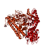
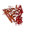
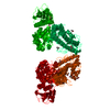
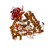
| Title | THE CRYSTAL STRUCTURE OF ALPHA-D-GLUCURONIDASE FROM BACILLUS STEAROTHERMOPHILUS T-1 COMPLEXED WITH GLUCURONIC ACID | ||||||
 Components Components | ALPHA-D-GLUCURONIDASE | ||||||
 Keywords Keywords | HYDROLASE | ||||||
| Function / homology |  Function and homology information Function and homology informationxylan alpha-1,2-glucuronosidase / xylan alpha-1,2-glucuronosidase activity / alpha-glucuronidase activity / glucuronoxylan catabolic process / extracellular region Similarity search - Function | ||||||
| Biological species |   Geobacillus stearothermophilus (bacteria) Geobacillus stearothermophilus (bacteria) | ||||||
| Method |  X-RAY DIFFRACTION / SYNCHROTRON / AB INITIO / Resolution: 1.65 Å X-RAY DIFFRACTION / SYNCHROTRON / AB INITIO / Resolution: 1.65 Å | ||||||
 Authors Authors | Golan, G. / Shallom, D. / Teplitsky, A. / Zaide, G. / Shulami, S. / Baasov, T. / Stojanoff, V. / Thompson, A. / Shoham, Y. / Shoham, G. | ||||||
 Citation Citation | Journal: J.Biol.Chem. / Year: 2004 Title: Crystal structures of Geobacillus stearothermophilus alpha-glucuronidase complexed with its substrate and products: mechanistic implications. Authors: Golan, G. / Shallom, D. / Teplitsky, A. / Zaide, G. / Shulami, S. / Baasov, T. / Stojanoff, V. / Thompson, A. / Shoham, Y. / Shoham, G. | ||||||
| History |
|
- Structure visualization
| Structure viewer | Molecule: MolmilJmol/JSmol |
|---|
- Downloads & links
-Download
| PDBx/mmCIF format | 1mqq.cif.gz | 168.2 KB | Display | PDBx/mmCIF format |
|---|---|---|---|---|
| PDB format | pdb1mqq.ent.gz | 133.1 KB | Display | PDB format |
| PDBx/mmJSON format | 1mqq.json.gz | Tree view | PDBx/mmJSON format | |
| Others |  Other downloads Other downloads |
-Validation report
| Arichive directory | https://data.pdbj.org/pub/pdb/validation_reports/mq/1mqqftp://data.pdbj.org/pub/pdb/validation_reports/mq/1mqq | HTTPS FTP |
|---|
-Related structure data
| Related structure data |  1k9dSC  1k9eC  1k9fC  1l8nC  1mqpC  1mqrC C: citing same article ( S: Starting model for refinement |
|---|---|
| Similar structure data |


-Links
 PDBj
PDBj
- Assembly
| Deposited unit | 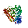
| |||||||||
|---|---|---|---|---|---|---|---|---|---|---|
| 1 | 
| |||||||||
| Unit cell |
| |||||||||
| Components on special symmetry positions |
|
-Components
| #1: Protein | Mass: 78579.234 Da / Num. of mol.: 1 Source method: isolated from a genetically manipulated source Source: (gene. exp.) Geobacillus stearothermophilus (bacteria)Strain: T-1 / Gene: AGUA / Plasmid: PET9D / Species (production host): Escherichia coli / Production host: | ||
|---|---|---|---|
| #2: Sugar | ChemComp-GCU /   Type: D-saccharide, alpha linking / Mass: 194.139 Da / Num. of mol.: 1 Type: D-saccharide, alpha linking / Mass: 194.139 Da / Num. of mol.: 1Source method: isolated from a genetically manipulated source Formula: C6H10O7 | ||
| #3: Chemical | ChemComp-GOL /   Mass: 92.094 Da / Num. of mol.: 11 / Source method: obtained synthetically / Formula: C3H8O3 Mass: 92.094 Da / Num. of mol.: 11 / Source method: obtained synthetically / Formula: C3H8O3#4: Water | ChemComp-HOH / |  Mass: 18.015 Da / Num. of mol.: 656 / Source method: isolated from a natural source / Formula: H2O Mass: 18.015 Da / Num. of mol.: 656 / Source method: isolated from a natural source / Formula: H2O |
-Experimental details
-Experiment
| Experiment | Method: X-RAY DIFFRACTION / Number of used crystals: 1 |
|---|
- Sample preparation
| Crystal | Density Matthews: 2.88 Å3/Da / Density % sol: 57.54 % |
|---|---|
| Crystal grow | Temperature: 293 K / Method: vapor diffusion, hanging drop / pH: 5 Details: 12% isopropanol, 14% PEG 4K, 0.1M Na-citrate, pH 5.0, VAPOR DIFFUSION, HANGING DROP, temperature 293K |
-Data collection
| Diffraction | Mean temperature: 95 K |
|---|---|
| Diffraction source | Source: SYNCHROTRON / Site: NSLS  / Beamline: X25 / Wavelength: 1.1 Å / Beamline: X25 / Wavelength: 1.1 Å |
| Detector | Type: BRANDEIS - B4 / Detector: CCD / Date: Jul 12, 2001 |
| Radiation | Protocol: SINGLE WAVELENGTH / Monochromatic (M) / Laue (L): M / Scattering type: x-ray |
| Radiation wavelength | Wavelength: 1.1 Å / Relative weight: 1 |
| Reflection | Resolution: 1.65→40 Å / Num. obs: 111221 / % possible obs: 98.9 % / Redundancy: 10 % / Rmerge(I) obs: 0.079 / Net I/σ(I): 7.9 |
| Reflection shell | Resolution: 1.65→1.68 Å / Redundancy: 10 % / Rmerge(I) obs: 0.452 / Num. unique all: 4995 / % possible all: 91 |
- Processing
| Software |
| |||||||||||||||||||||||||||||||||
|---|---|---|---|---|---|---|---|---|---|---|---|---|---|---|---|---|---|---|---|---|---|---|---|---|---|---|---|---|---|---|---|---|---|---|
| Refinement | Method to determine structure: AB INITIO Starting model: PDB CODE 1K9D Resolution: 1.65→40 Å / Num. parameters: 25802 / Num. restraintsaints: 24044 / Cross valid method: FREE R / σ(F): 0 / Stereochemistry target values: ENGH AND HUBER
| |||||||||||||||||||||||||||||||||
| Refine analyze | Luzzati coordinate error obs: 0.15 Å / Num. disordered residues: 44 / Occupancy sum hydrogen: 0 / Occupancy sum non hydrogen: 6237.05 | |||||||||||||||||||||||||||||||||
| Refinement step | Cycle: LAST / Resolution: 1.65→40 Å
| |||||||||||||||||||||||||||||||||
| Refine LS restraints |
|
