 Movie
Movie Controller

[English] 日本語
 Yorodumi
- PDB-1k9e: Crystal structure of a mutated family-67 alpha-D-glucuronidase (E... -
+ Open data

- Basic information
| Entry | Database: PDB / ID: 1k9e | ||||||
|---|---|---|---|---|---|---|---|
| Title | Crystal structure of a mutated family-67 alpha-D-glucuronidase (E285N) from Bacillus stearothermophilus T-6, complexed with 4-O-methyl-glucuronic acid | ||||||
 Components Components | alpha-D-glucuronidase | ||||||
 Keywords Keywords | HYDROLASE | ||||||
| Function / homology |  Function and homology information Function and homology informationxylan alpha-1,2-glucuronosidase / xylan alpha-1,2-glucuronosidase activity / alpha-glucuronidase activity / glucuronoxylan catabolic process / Hydrolases; Glycosylases; Glycosidases, i.e. enzymes that hydrolyse O- and S-glycosyl compounds / extracellular region Similarity search - Function | ||||||
| Biological species |   Geobacillus stearothermophilus (bacteria) Geobacillus stearothermophilus (bacteria) | ||||||
| Method |  X-RAY DIFFRACTION / SYNCHROTRON / AB INITIO PHASING / Resolution: 1.85 Å X-RAY DIFFRACTION / SYNCHROTRON / AB INITIO PHASING / Resolution: 1.85 Å | ||||||
 Authors Authors | Golan, G. / Shallom, D. / Teplitsky, A. / Zaide, G. / Shulami, S. / Baasov, T. / Stojanoff, V. / Thompson, A. / Shoham, Y. / Shoham, G. | ||||||
 Citation Citation | Journal: J.Biol.Chem. / Year: 2004 Title: Crystal Structures of Geobacillus stearothermophilus {alpha}-Glucuronidase Complexed with Its Substrate and Products: MECHANISTIC IMPLICATIONS. Authors: Golan, G. / Shallom, D. / Teplitsky, A. / Zaide, G. / Shulami, S. / Baasov, T. / Stojanoff, V. / Thompson, A. / Shoham, Y. / Shoham, G. | ||||||
| History |
|
- Structure visualization
| Structure viewer | Molecule: MolmilJmol/JSmol |
|---|
- Downloads & links
-Download
| PDBx/mmCIF format | 1k9e.cif.gz | 160.6 KB | Display | PDBx/mmCIF format |
|---|---|---|---|---|
| PDB format | pdb1k9e.ent.gz | 125.8 KB | Display | PDB format |
| PDBx/mmJSON format | 1k9e.json.gz | Tree view | PDBx/mmJSON format | |
| Others |  Other downloads Other downloads |
-Validation report
| Arichive directory | https://data.pdbj.org/pub/pdb/validation_reports/k9/1k9eftp://data.pdbj.org/pub/pdb/validation_reports/k9/1k9e | HTTPS FTP |
|---|
-Related structure data
| Related structure data |  1k9dSC  1k9fC  1l8nC  1mqpC  1mqqC  1mqrC S: Starting model for refinement C: citing same article ( |
|---|---|
| Similar structure data |


-Links
 PDBj
PDBj
- Assembly
| Deposited unit | 
| ||||||||
|---|---|---|---|---|---|---|---|---|---|
| 1 | 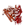
| ||||||||
| Unit cell |
|
-Components
| #1: Protein | Mass: 78563.258 Da / Num. of mol.: 1 / Mutation: E285N Source method: isolated from a genetically manipulated source Source: (gene. exp.) Geobacillus stearothermophilus (bacteria)Strain: T6 / Gene: AGUA / Plasmid: pET9d / Species (production host): Escherichia coli / Production host: References: UniProt: Q8VVD2, UniProt: Q09LY5*PLUS, EC: 3.2.1.139 | ||
|---|---|---|---|
| #2: Sugar | ChemComp-GCV /   Type: D-saccharide, alpha linking / Mass: 208.166 Da / Num. of mol.: 1 Type: D-saccharide, alpha linking / Mass: 208.166 Da / Num. of mol.: 1Source method: isolated from a genetically manipulated source Formula: C7H12O7 | ||
| #3: Chemical | ChemComp-GOL /   Mass: 92.094 Da / Num. of mol.: 11 / Source method: obtained synthetically / Formula: C3H8O3 Mass: 92.094 Da / Num. of mol.: 11 / Source method: obtained synthetically / Formula: C3H8O3#4: Water | ChemComp-HOH / |  Mass: 18.015 Da / Num. of mol.: 483 / Source method: isolated from a natural source / Formula: H2O Mass: 18.015 Da / Num. of mol.: 483 / Source method: isolated from a natural source / Formula: H2O |
-Experimental details
-Experiment
| Experiment | Method: X-RAY DIFFRACTION / Number of used crystals: 1 |
|---|
- Sample preparation
| Crystal | Density Matthews: 2.85 Å3/Da / Density % sol: 56.78 % | ||||||||||||||||||||||||||||||
|---|---|---|---|---|---|---|---|---|---|---|---|---|---|---|---|---|---|---|---|---|---|---|---|---|---|---|---|---|---|---|---|
| Crystal grow | Temperature: 293 K / Method: vapor diffusion, hanging drop / pH: 5 Details: 0.1M Na-citrate pH 5.0, 14% PEG 4000, 12% Iso-propanol, VAPOR DIFFUSION, HANGING DROP, temperature 293K | ||||||||||||||||||||||||||||||
| Crystal grow | *PLUS Temperature: 20 ℃ / PH range low: 5.5 / PH range high: 5 | ||||||||||||||||||||||||||||||
| Components of the solutions | *PLUS
|
-Data collection
| Diffraction | Mean temperature: 95 K |
|---|---|
| Diffraction source | Source: SYNCHROTRON / Site: NSLS  / Beamline: X12B / Wavelength: 0.9766 Å / Beamline: X12B / Wavelength: 0.9766 Å |
| Detector | Type: ADSC QUANTUM 4 / Detector: CCD / Date: Jul 29, 2000 |
| Radiation | Protocol: SINGLE WAVELENGTH / Monochromatic (M) / Laue (L): M / Scattering type: x-ray |
| Radiation wavelength | Wavelength: 0.9766 Å / Relative weight: 1 |
| Reflection | Resolution: 1.85→40 Å / Num. obs: 72670 / % possible obs: 92.1 % / Observed criterion σ(I): 1 / Redundancy: 6 % / Biso Wilson estimate: 15 Å2 / Rmerge(I) obs: 0.069 / Net I/σ(I): 12.6 |
| Reflection shell | Resolution: 1.85→1.88 Å / Rmerge(I) obs: 0.234 / % possible all: 63.1 |
| Reflection | *PLUS Lowest resolution: 40 Å / Num. measured all: 354446 |
| Reflection shell | *PLUS % possible obs: 63.1 % |
- Processing
| Software |
| ||||||||||||||||||||||||||||||||||||
|---|---|---|---|---|---|---|---|---|---|---|---|---|---|---|---|---|---|---|---|---|---|---|---|---|---|---|---|---|---|---|---|---|---|---|---|---|---|
| Refinement | Method to determine structure: AB INITIO PHASING Starting model: PDB ENTRY 1K9D Resolution: 1.85→36.03 Å / Rfactor Rfree error: 0.003 / Data cutoff high absF: 192936.65 / Data cutoff low absF: 0 / Isotropic thermal model: RESTRAINED / Cross valid method: THROUGHOUT / σ(F): 1
| ||||||||||||||||||||||||||||||||||||
| Solvent computation | Solvent model: FLAT MODEL / Bsol: 45.0392 Å2 / ksol: 0.374165 e/Å3 | ||||||||||||||||||||||||||||||||||||
| Displacement parameters | Biso mean: 23.5 Å2
| ||||||||||||||||||||||||||||||||||||
| Refine analyze |
| ||||||||||||||||||||||||||||||||||||
| Refinement step | Cycle: LAST / Resolution: 1.85→36.03 Å
| ||||||||||||||||||||||||||||||||||||
| Refine LS restraints |
| ||||||||||||||||||||||||||||||||||||
| LS refinement shell | Resolution: 1.85→1.97 Å / Rfactor Rfree error: 0.01 / Total num. of bins used: 6
| ||||||||||||||||||||||||||||||||||||
| Xplor file |
| ||||||||||||||||||||||||||||||||||||
| Refine LS restraints | *PLUS
|
