 Movie
Movie Controller

[English] 日本語
 Yorodumi
- PDB-1l8n: The 1.5A crystal structure of alpha-D-glucuronidase from Bacillus... -
+ Open data

- Basic information
| Entry | Database: PDB / ID: 1l8n | |||||||||
|---|---|---|---|---|---|---|---|---|---|---|
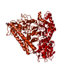
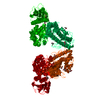
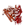
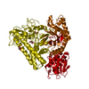
| Title | The 1.5A crystal structure of alpha-D-glucuronidase from Bacillus stearothermophilus T-1, complexed with 4-O-methyl-glucuronic acid and xylotriose | |||||||||
 Components Components | ALPHA-D-GLUCURONIDASE | |||||||||
 Keywords Keywords | HYDROLASE | |||||||||
| Function / homology |  Function and homology information Function and homology informationxylan alpha-1,2-glucuronosidase / xylan alpha-1,2-glucuronosidase activity / alpha-glucuronidase activity / glucuronoxylan catabolic process / extracellular region Similarity search - Function | |||||||||
| Biological species |   Geobacillus stearothermophilus (bacteria) Geobacillus stearothermophilus (bacteria) | |||||||||
| Method |  X-RAY DIFFRACTION / SYNCHROTRON / AB INITIO / Resolution: 1.5 Å X-RAY DIFFRACTION / SYNCHROTRON / AB INITIO / Resolution: 1.5 Å | |||||||||
 Authors Authors | Golan, G. / Shallom, D. / Teplitsky, A. / Zaide, G. / Shulami, S. / Baasov, T. / Stojanoff, V. / Thompson, A. / Shoham, Y. / Shoham, G. | |||||||||
 Citation Citation | Journal: J.Biol.Chem. / Year: 2004 Title: Crystal Structures of Geobacillus stearothermophilus {alpha}-Glucuronidase Complexed with Its Substrate and Products: MECHANISTIC IMPLICATIONS. Authors: Golan, G. / Shallom, D. / Teplitsky, A. / Zaide, G. / Shulami, S. / Baasov, T. / Stojanoff, V. / Thompson, A. / Shoham, Y. / Shoham, G. | |||||||||
| History |
| |||||||||
| Remark 600 | HETEROGEN THE PRODUCT IS XYLOTRIOSE. IN THE ENTRY, XYP 702 AND XYP 703 ARE COVALENTLY BOUND. THE ...HETEROGEN THE PRODUCT IS XYLOTRIOSE. IN THE ENTRY, XYP 702 AND XYP 703 ARE COVALENTLY BOUND. THE THIRD XYLOSE IS NOT MODELED DUE TO LACK OF FIXED POSITION AND CLEAR DENSITY. THE BOND BETWEEN GCW 701 AND XYP 702 WAS BROKEN BY THE ENZYME. ALSO, XYP 702 AND GOL 763 OCCUPY THE SAME SPACE AND EACH HAVE HALF OCCUPANCY. |
- Structure visualization
| Structure viewer | Molecule: MolmilJmol/JSmol |
|---|
- Downloads & links
-Download
| PDBx/mmCIF format | 1l8n.cif.gz | 321.2 KB | Display | PDBx/mmCIF format |
|---|---|---|---|---|
| PDB format | pdb1l8n.ent.gz | 260 KB | Display | PDB format |
| PDBx/mmJSON format | 1l8n.json.gz | Tree view | PDBx/mmJSON format | |
| Others |  Other downloads Other downloads |
-Validation report
| Arichive directory | https://data.pdbj.org/pub/pdb/validation_reports/l8/1l8nftp://data.pdbj.org/pub/pdb/validation_reports/l8/1l8n | HTTPS FTP |
|---|
-Related structure data
| Related structure data |  1k9dSC  1k9eC  1k9fC  1mqpC  1mqqC  1mqrC C: citing same article ( S: Starting model for refinement |
|---|---|
| Similar structure data |


-Links
 PDBj
PDBj
- Assembly
| Deposited unit | 
| |||||||||
|---|---|---|---|---|---|---|---|---|---|---|
| 1 | 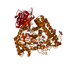
| |||||||||
| Unit cell |
| |||||||||
| Components on special symmetry positions |
|
-Components
| #1: Protein | Mass: 78579.234 Da / Num. of mol.: 1 Source method: isolated from a genetically manipulated source Source: (gene. exp.) Geobacillus stearothermophilus (bacteria)Gene: AGUA / Plasmid: PET9D / Species (production host): Escherichia coli / Production host: References: GenBank: 16930794, UniProt: Q8VVD2*PLUS, EC: 3.2.1.139 | ||
|---|---|---|---|
| #2: Polysaccharide | beta-D-xylopyranose-(1-4)-beta-D-xylopyranose / 4beta-beta-xylobiose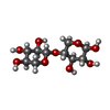  Source method: isolated from a genetically manipulated source Details: oligosaccharide / References: 4beta-beta-xylobiose | ||
| #3: Sugar | ChemComp-GCW /   Type: D-saccharide, beta linking / Mass: 208.166 Da / Num. of mol.: 1 Type: D-saccharide, beta linking / Mass: 208.166 Da / Num. of mol.: 1Source method: isolated from a genetically manipulated source Formula: C7H12O7 | ||
| #4: Chemical | ChemComp-GOL /   Mass: 92.094 Da / Num. of mol.: 14 / Source method: obtained synthetically / Formula: C3H8O3 Mass: 92.094 Da / Num. of mol.: 14 / Source method: obtained synthetically / Formula: C3H8O3#5: Water | ChemComp-HOH / |  Mass: 18.015 Da / Num. of mol.: 667 / Source method: isolated from a natural source / Formula: H2O Mass: 18.015 Da / Num. of mol.: 667 / Source method: isolated from a natural source / Formula: H2O |
-Experimental details
-Experiment
| Experiment | Method: X-RAY DIFFRACTION / Number of used crystals: 1 |
|---|
- Sample preparation
| Crystal | Density Matthews: 2.53 Å3/Da / Density % sol: 51.3 % |
|---|---|
| Crystal grow | Temperature: 293 K / Method: vapor diffusion, hanging drop / pH: 5 Details: 0.1M NA-CITRATE PH 5.0, 14% PEG 4000, 12% ISO-PROPANOL, VAPOR DIFFUSION, HANGING DROP, temperature 293K |
-Data collection
| Diffraction | Mean temperature: 95 K |
|---|---|
| Diffraction source | Source: SYNCHROTRON / Site: NSLS  / Beamline: X25 / Wavelength: 1.1 Å / Beamline: X25 / Wavelength: 1.1 Å |
| Detector | Type: BRANDEIS / Detector: CCD / Date: Jul 12, 2001 |
| Radiation | Protocol: SINGLE WAVELENGTH / Monochromatic (M) / Laue (L): M / Scattering type: x-ray |
| Radiation wavelength | Wavelength: 1.1 Å / Relative weight: 1 |
| Reflection | Resolution: 1.5→40 Å / Num. obs: 145976 / % possible obs: 98.8 % / Observed criterion σ(I): -3 / Redundancy: 15 % / Rmerge(I) obs: 0.11 / Net I/σ(I): 6.8 |
| Reflection shell | Resolution: 1.5→1.53 Å / Rmerge(I) obs: 0.399 / Num. unique all: 6525 / % possible all: 90.3 |
- Processing
| Software |
| |||||||||||||||||||||||||
|---|---|---|---|---|---|---|---|---|---|---|---|---|---|---|---|---|---|---|---|---|---|---|---|---|---|---|
| Refinement | Method to determine structure: AB INITIO Starting model: pdb code 1k9d Resolution: 1.5→10 Å / Cross valid method: FREE R / σ(F): 0 / Stereochemistry target values: ENGH & HUBER
| |||||||||||||||||||||||||
| Refine analyze | Num. disordered residues: 42 | |||||||||||||||||||||||||
| Refinement step | Cycle: LAST / Resolution: 1.5→10 Å
| |||||||||||||||||||||||||
| Refine LS restraints |
|
