 Movie
Movie Controller

[English] 日本語
 Yorodumi
- PDB-1nd5: Crystal Structures of Human Prostatic Acid Phosphatase in Complex... -
+ Open data

- Basic information
| Entry | Database: PDB / ID: 1nd5 | ||||||||||||
|---|---|---|---|---|---|---|---|---|---|---|---|---|---|
| Title | Crystal Structures of Human Prostatic Acid Phosphatase in Complex with a Phosphate Ion and alpha-Benzylaminobenzylphosphonic Acid Update the Mechanistic Picture and Offer New Insights into Inhibitor Design | ||||||||||||
 Components Components | prostatic acid phosphatase | ||||||||||||
 Keywords Keywords | HYDROLASE / Acid Phosphatase / PAP / Prostate / Phosphate / inhibitor | ||||||||||||
| Function / homology |  Function and homology information Function and homology informationthiamine phosphate phosphatase activity / positive regulation of adenosine receptor signaling pathway / thiamine metabolic process / Golgi cisterna / adenosine metabolic process / acid phosphatase / acid phosphatase activity / regulation of sensory perception of pain / lysophosphatidic acid phosphatase activity / 5'-nucleotidase ...thiamine phosphate phosphatase activity / positive regulation of adenosine receptor signaling pathway / thiamine metabolic process / Golgi cisterna / adenosine metabolic process / acid phosphatase / acid phosphatase activity / regulation of sensory perception of pain / lysophosphatidic acid phosphatase activity / 5'-nucleotidase / choline binding / 5'-nucleotidase activity / nucleotide metabolic process / vesicle membrane / azurophil granule membrane / purine nucleobase metabolic process / phosphatase activity / multivesicular body / protein-tyrosine-phosphatase / protein tyrosine phosphatase activity / filopodium / lipid metabolic process / apical part of cell / molecular adaptor activity / lysosomal membrane / Neutrophil degranulation / protein homodimerization activity / : / extracellular exosome / identical protein binding / nucleus / plasma membrane / cytosol Similarity search - Function | ||||||||||||
| Biological species |  Homo sapiens (human) Homo sapiens (human) | ||||||||||||
| Method |  X-RAY DIFFRACTION / FOURIER SYNTHESIS / Resolution: 2.9 Å X-RAY DIFFRACTION / FOURIER SYNTHESIS / Resolution: 2.9 Å | ||||||||||||
 Authors Authors | Ortlund, E. / LaCount, M.W. / Lebioda, L. | ||||||||||||
 Citation Citation | Journal: Biochemistry / Year: 2003 Title: Crystal structures of human prostatic acid phosphatase in complex with a phosphate ion and alpha-benzylaminobenzylphosphonic acid update the mechanistic picture and offer new insights into inhibitor design Authors: Ortlund, E. / LaCount, M.W. / Lebioda, L. | ||||||||||||
| History |
|
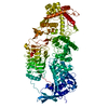
- Structure visualization
| Structure viewer | Molecule: MolmilJmol/JSmol |
|---|
- Downloads & links
-Download
| PDBx/mmCIF format | 1nd5.cif.gz | 298.8 KB | Display | PDBx/mmCIF format |
|---|---|---|---|---|
| PDB format | pdb1nd5.ent.gz | 242.3 KB | Display | PDB format |
| PDBx/mmJSON format | 1nd5.json.gz | Tree view | PDBx/mmJSON format | |
| Others |  Other downloads Other downloads |
-Validation report
| Arichive directory | https://data.pdbj.org/pub/pdb/validation_reports/nd/1nd5ftp://data.pdbj.org/pub/pdb/validation_reports/nd/1nd5 | HTTPS FTP |
|---|
-Related structure data
| Related structure data |  1nd6C  2hpaS S: Starting model for refinement C: citing same article ( |
|---|---|
| Similar structure data |


-Links
 PDBj
PDBj


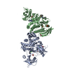
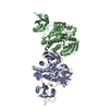
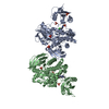
- Assembly
| Deposited unit | 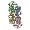
| ||||||||||
|---|---|---|---|---|---|---|---|---|---|---|---|
| 1 | 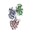
| ||||||||||
| 2 | 
| ||||||||||
| Unit cell |
| ||||||||||
| Details | The Biological Assembley is a dimer. The asymmetric unit contains two such dimers. |
-Components
-Protein , 1 types, 4 molecules ABCD
| #1: Protein | Mass: 41050.602 Da / Num. of mol.: 4 / Source method: isolated from a natural source / Source: (natural) Homo sapiens (human) / Tissue: semen / Tissue fraction: Seminal Plasma / References: UniProt: P15309, acid phosphatase |
|---|
-Sugars , 4 types, 7 molecules 

| #2: Polysaccharide | 2-acetamido-2-deoxy-beta-D-glucopyranose-(1-4)-2-acetamido-2-deoxy-beta-D-glucopyranose Source method: isolated from a genetically manipulated source #3: Polysaccharide | alpha-D-mannopyranose-(1-4)-2-acetamido-2-deoxy-beta-D-glucopyranose-(1-4)-2-acetamido-2-deoxy-beta- ...alpha-D-mannopyranose-(1-4)-2-acetamido-2-deoxy-beta-D-glucopyranose-(1-4)-2-acetamido-2-deoxy-beta-D-glucopyranose | Source method: isolated from a genetically manipulated source #6: Sugar | ChemComp-NDG / |  Type: D-saccharide, alpha linking / Mass: 221.208 Da / Num. of mol.: 1 Type: D-saccharide, alpha linking / Mass: 221.208 Da / Num. of mol.: 1Source method: isolated from a genetically manipulated source Formula: C8H15NO6 #7: Sugar | ChemComp-NAG / |  Type: D-saccharide, beta linking / Mass: 221.208 Da / Num. of mol.: 1 Type: D-saccharide, beta linking / Mass: 221.208 Da / Num. of mol.: 1Source method: isolated from a genetically manipulated source Formula: C8H15NO6 |
|---|
-Non-polymers , 3 types, 147 molecules 


| #4: Chemical | ChemComp-2BF /  Mass: 277.256 Da / Num. of mol.: 4 / Source method: obtained synthetically / Formula: C14H16NO3P Mass: 277.256 Da / Num. of mol.: 4 / Source method: obtained synthetically / Formula: C14H16NO3P#5: Chemical | ChemComp-1PE /  Mass: 238.278 Da / Num. of mol.: 8 / Source method: obtained synthetically / Formula: C10H22O6 / Comment: precipitant*YM Mass: 238.278 Da / Num. of mol.: 8 / Source method: obtained synthetically / Formula: C10H22O6 / Comment: precipitant*YM#8: Water | ChemComp-HOH / | Mass: 18.015 Da / Num. of mol.: 135 / Source method: isolated from a natural source / Formula: H2O |
|---|
-Details
| Has protein modification | Y |
|---|
-Experimental details
-Experiment
| Experiment | Method: X-RAY DIFFRACTION / Number of used crystals: 1 |
|---|
- Sample preparation
| Crystal | Density Matthews: 2.67 Å3/Da / Density % sol: 53.87 % | |||||||||||||||||||||||||||||||||||||||||||||||||
|---|---|---|---|---|---|---|---|---|---|---|---|---|---|---|---|---|---|---|---|---|---|---|---|---|---|---|---|---|---|---|---|---|---|---|---|---|---|---|---|---|---|---|---|---|---|---|---|---|---|---|
| Crystal grow | Temperature: 298 K / Method: vapor diffusion, hanging drop / pH: 10 Details: PEG, KCL, Glycine, pH 10.0, VAPOR DIFFUSION, HANGING DROP, temperature 298K | |||||||||||||||||||||||||||||||||||||||||||||||||
| Crystal grow | *PLUS Method: unknown | |||||||||||||||||||||||||||||||||||||||||||||||||
| Components of the solutions | *PLUS
|
-Data collection
| Diffraction | Mean temperature: 298 K |
|---|---|
| Diffraction source | Source: ROTATING ANODE / Type: RIGAKU RU200 / Wavelength: 1.5418 Å |
| Detector | Type: RIGAKU RAXIS IV / Detector: IMAGE PLATE / Date: Dec 1, 1997 / Details: Yale Mirrors |
| Radiation | Monochromator: Yale Mirrors / Protocol: SINGLE WAVELENGTH / Monochromatic (M) / Laue (L): M / Scattering type: x-ray |
| Radiation wavelength | Wavelength: 1.5418 Å / Relative weight: 1 |
| Reflection | Resolution: 2.89→50 Å / Num. all: 33191 / Num. obs: 33191 / % possible obs: 82.2 % / Biso Wilson estimate: 74.4 Å2 / Rmerge(I) obs: 0.063 |
| Reflection shell | Resolution: 2.89→3 Å / % possible all: 63.7 |
| Reflection | *PLUS Highest resolution: 2.9 Å / Lowest resolution: 50 Å |
| Reflection shell | *PLUS Lowest resolution: 3 Å / % possible obs: 63.7 % |
- Processing
| Software |
| |||||||||||||||||||||||||
|---|---|---|---|---|---|---|---|---|---|---|---|---|---|---|---|---|---|---|---|---|---|---|---|---|---|---|
| Refinement | Method to determine structure: FOURIER SYNTHESIS Starting model: 2hpa Resolution: 2.9→77.94 Å / Rfactor Rfree error: 0.007 / Data cutoff high absF: 245501.33 / Data cutoff low absF: 0 / Isotropic thermal model: RESTRAINED / Cross valid method: THROUGHOUT / σ(F): 0 / Stereochemistry target values: Engh & Huber
| |||||||||||||||||||||||||
| Solvent computation | Solvent model: FLAT MODEL / Bsol: 34.2524 Å2 / ksol: 0.354636 e/Å3 | |||||||||||||||||||||||||
| Displacement parameters | Biso mean: 30 Å2
| |||||||||||||||||||||||||
| Refine analyze | Luzzati coordinate error free: 0.46 Å / Luzzati sigma a free: 0.5 Å | |||||||||||||||||||||||||
| Refinement step | Cycle: LAST / Resolution: 2.9→77.94 Å
| |||||||||||||||||||||||||
| Refine LS restraints |
| |||||||||||||||||||||||||
| LS refinement shell | Resolution: 2.9→3.08 Å / Rfactor Rfree error: 0.025 / Total num. of bins used: 6
| |||||||||||||||||||||||||
| Xplor file |
| |||||||||||||||||||||||||
| Refinement | *PLUS Highest resolution: 2.9 Å / Lowest resolution: 50 Å / Rfactor Rfree: 0.278 / Rfactor Rwork: 0.202 | |||||||||||||||||||||||||
| Solvent computation | *PLUS | |||||||||||||||||||||||||
| Displacement parameters | *PLUS | |||||||||||||||||||||||||
| Refine LS restraints | *PLUS
|
