 Movie
Movie Controller

[English] 日本語
 Yorodumi
- PDB-2hpa: STRUCTURAL ORIGINS OF L(+)-TARTRATE INHIBITION OF HUMAN PROSTATIC... -
+ Open data

- Basic information
| Entry | Database: PDB / ID: 2hpa | ||||||||||||
|---|---|---|---|---|---|---|---|---|---|---|---|---|---|
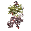
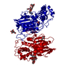
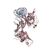
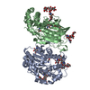
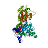
| Title | STRUCTURAL ORIGINS OF L(+)-TARTRATE INHIBITION OF HUMAN PROSTATIC ACID PHOSPHATASE | ||||||||||||
 Components Components | PROTEIN (ACID PHOSPHATASE) | ||||||||||||
 Keywords Keywords | HYDROLASE / ACID PHOSPHATASE / N-PROPYLTARTRAMATE | ||||||||||||
| Function / homology |  Function and homology information Function and homology informationthiamine phosphate phosphatase activity / positive regulation of adenosine receptor signaling pathway / thiamine metabolic process / Golgi cisterna / adenosine metabolic process / acid phosphatase / acid phosphatase activity / regulation of sensory perception of pain / lysophosphatidic acid phosphatase activity / 5'-nucleotidase ...thiamine phosphate phosphatase activity / positive regulation of adenosine receptor signaling pathway / thiamine metabolic process / Golgi cisterna / adenosine metabolic process / acid phosphatase / acid phosphatase activity / regulation of sensory perception of pain / lysophosphatidic acid phosphatase activity / 5'-nucleotidase / choline binding / 5'-nucleotidase activity / nucleotide metabolic process / vesicle membrane / azurophil granule membrane / purine nucleobase metabolic process / phosphatase activity / multivesicular body / protein-tyrosine-phosphatase / protein tyrosine phosphatase activity / filopodium / lipid metabolic process / apical part of cell / molecular adaptor activity / lysosomal membrane / Neutrophil degranulation / protein homodimerization activity / : / extracellular exosome / identical protein binding / nucleus / plasma membrane / cytosol Similarity search - Function | ||||||||||||
| Biological species |  Homo sapiens (human) Homo sapiens (human) | ||||||||||||
| Method |  X-RAY DIFFRACTION / OTHER / Resolution: 2.9 Å X-RAY DIFFRACTION / OTHER / Resolution: 2.9 Å | ||||||||||||
 Authors Authors | Lacount, M.W. / Handy, G. / Lebioda, L. | ||||||||||||
 Citation Citation | Journal: J.Biol.Chem. / Year: 1998 Title: Structural origins of L(+)-tartrate inhibition of human prostatic acid phosphatase. Authors: LaCount, M.W. / Handy, G. / Lebioda, L. #1: Journal: J.Biol.Chem. / Year: 1993Title: Three-Dimensional Structure of Rat Acid Phosphatase in Complex with L(+)-Tartrate Authors: Lindqvist, Y. / Schneider, G. / Vihko, P. | ||||||||||||
| History |
|
- Structure visualization
| Structure viewer | Molecule: MolmilJmol/JSmol |
|---|
- Downloads & links
-Download
| PDBx/mmCIF format | 2hpa.cif.gz | 297.7 KB | Display | PDBx/mmCIF format |
|---|---|---|---|---|
| PDB format | pdb2hpa.ent.gz | 240.4 KB | Display | PDB format |
| PDBx/mmJSON format | 2hpa.json.gz | Tree view | PDBx/mmJSON format | |
| Others |  Other downloads Other downloads |
-Validation report
| Arichive directory | https://data.pdbj.org/pub/pdb/validation_reports/hp/2hpaftp://data.pdbj.org/pub/pdb/validation_reports/hp/2hpa | HTTPS FTP |
|---|
-Related structure data
| Similar structure data |
|---|


-Links
 PDBj
PDBj


- Assembly
| Deposited unit | 
| ||||||||
|---|---|---|---|---|---|---|---|---|---|
| 1 | 
| ||||||||
| 2 | 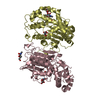
| ||||||||
| 3 | 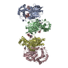
| ||||||||
| Unit cell |
| ||||||||
| Noncrystallographic symmetry (NCS) | NCS oper: (Code: given Matrix: (-0.792218, 0.090391, -0.603507), Vector: |
-Components
-Protein , 1 types, 4 molecules ABCD
| #1: Protein | Mass: 39776.426 Da / Num. of mol.: 4 / Source method: isolated from a natural source / Details: ISOLATED FROM HUMAN SEMINAL FLUID / Source: (natural) Homo sapiens (human) / References: UniProt: P15309, acid phosphatase |
|---|
-Sugars , 4 types, 8 molecules 
| #2: Polysaccharide | alpha-D-mannopyranose-(1-6)-beta-D-mannopyranose-(1-4)-2-acetamido-2-deoxy-beta-D-glucopyranose Source method: isolated from a genetically manipulated source |
|---|---|
| #3: Polysaccharide | 2-acetamido-2-deoxy-beta-D-glucopyranose-(1-4)-2-acetamido-2-deoxy-beta-D-glucopyranose |
| #4: Polysaccharide | beta-D-mannopyranose-(1-4)-2-acetamido-2-deoxy-beta-D-glucopyranose-(1-4)-2-acetamido-2-deoxy-beta- ...beta-D-mannopyranose-(1-4)-2-acetamido-2-deoxy-beta-D-glucopyranose-(1-4)-2-acetamido-2-deoxy-beta-D-glucopyranose |
| #5: Sugar | ChemComp-NAG /  Type: D-saccharide, beta linking / Mass: 221.208 Da / Num. of mol.: 5 Type: D-saccharide, beta linking / Mass: 221.208 Da / Num. of mol.: 5Source method: isolated from a genetically manipulated source Formula: C8H15NO6 |
-Non-polymers , 2 types, 353 molecules 

| #6: Chemical |  Mass: 191.182 Da / Num. of mol.: 3 / Source method: obtained synthetically / Formula: C7H13NO5 Mass: 191.182 Da / Num. of mol.: 3 / Source method: obtained synthetically / Formula: C7H13NO5#7: Water | ChemComp-HOH / | Mass: 18.015 Da / Num. of mol.: 350 / Source method: isolated from a natural source / Formula: H2O |
|---|
-Details
| Has protein modification | Y |
|---|
-Experimental details
-Experiment
| Experiment | Method: X-RAY DIFFRACTION / Number of used crystals: 1 |
|---|
- Sample preparation
| Crystal | Density Matthews: 2.71 Å3/Da / Density % sol: 54.66 % | ||||||||||||||||||||||||||||||
|---|---|---|---|---|---|---|---|---|---|---|---|---|---|---|---|---|---|---|---|---|---|---|---|---|---|---|---|---|---|---|---|
| Crystal grow | pH: 10 / Details: pH 10 | ||||||||||||||||||||||||||||||
| Crystal grow | *PLUS Method: unknown | ||||||||||||||||||||||||||||||
| Components of the solutions | *PLUS
|
-Data collection
| Diffraction | Mean temperature: 123 K |
|---|---|
| Diffraction source | Source: ROTATING ANODE / Type: RIGAKU RU200 / Wavelength: 1.5418 |
| Detector | Type: RIGAKU RAXIS IV / Detector: IMAGE PLATE / Details: MIRRORS |
| Radiation | Monochromator: NI FILTER / Protocol: SINGLE WAVELENGTH / Monochromatic (M) / Laue (L): M / Scattering type: x-ray |
| Radiation wavelength | Wavelength: 1.5418 Å / Relative weight: 1 |
| Reflection | Resolution: 2.9→99 Å / Num. obs: 39548 / % possible obs: 98.5 % / Observed criterion σ(I): 1 / Redundancy: 5 % / Biso Wilson estimate: 72.3 Å2 / Rmerge(I) obs: 0.138 |
| Reflection | *PLUS Num. measured all: 224797 |
| Reflection shell | *PLUS Highest resolution: 2.88 Å / Lowest resolution: 2.98 Å / % possible obs: 60 % |
- Processing
| Software |
| ||||||||||||||||||||||||||||||||||||||||||||||||||||||||||||
|---|---|---|---|---|---|---|---|---|---|---|---|---|---|---|---|---|---|---|---|---|---|---|---|---|---|---|---|---|---|---|---|---|---|---|---|---|---|---|---|---|---|---|---|---|---|---|---|---|---|---|---|---|---|---|---|---|---|---|---|---|---|
| Refinement | Method to determine structure: OTHER / Resolution: 2.9→8 Å / Rfactor Rfree error: 0.008 / Data cutoff high absF: 10000000 / Data cutoff low absF: 0.001 / Isotropic thermal model: RESTRAINED / Cross valid method: THROUGHOUT / σ(F): 2
| ||||||||||||||||||||||||||||||||||||||||||||||||||||||||||||
| Displacement parameters | Biso mean: 17.5 Å2
| ||||||||||||||||||||||||||||||||||||||||||||||||||||||||||||
| Refine analyze |
| ||||||||||||||||||||||||||||||||||||||||||||||||||||||||||||
| Refinement step | Cycle: LAST / Resolution: 2.9→8 Å
| ||||||||||||||||||||||||||||||||||||||||||||||||||||||||||||
| Refine LS restraints |
| ||||||||||||||||||||||||||||||||||||||||||||||||||||||||||||
| Refine LS restraints NCS | NCS model details: RESTRAIN | ||||||||||||||||||||||||||||||||||||||||||||||||||||||||||||
| LS refinement shell | Resolution: 2.9→3.07 Å / Rfactor Rfree error: 0.025 / Total num. of bins used: 6
| ||||||||||||||||||||||||||||||||||||||||||||||||||||||||||||
| Xplor file |
| ||||||||||||||||||||||||||||||||||||||||||||||||||||||||||||
| Software | *PLUS Name: X-PLOR / Version: 3.851 / Classification: refinement | ||||||||||||||||||||||||||||||||||||||||||||||||||||||||||||
| Refine LS restraints | *PLUS
|
