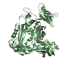
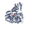
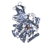
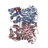
- PDB-5vbn: Crystal Structure of human DNA polymerase epsilon B-subunit in co... -
+
Open data
ID or keywords:
Loading...
-
Basic information
Entry
Database: PDB / ID: 5vbn
Title
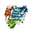
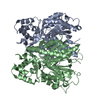
Crystal Structure of human DNA polymerase epsilon B-subunit in complex with C-terminal domain of catalytic subunit
Components
DNA polymerase epsilon catalytic subunit A
DNA polymerase epsilon subunit 2
Keywords
TRANSFERASE / replication / DNA replication / polymerase / DNA polymerase / DNA polymerase epsilon / B-subunit / catalytic subunit
Function / homology
Function and homology information
DNA replication initiation / epsilon DNA polymerase complex / nucleotide-excision repair, DNA gap filling / DNA replication proofreading / single-stranded DNA 3'-5' DNA exonuclease activity / Hydrolases; Acting on ester bonds; Exodeoxyribonucleases producing 5'-phosphomonoesters / leading strand elongation / DNA synthesis involved in DNA repair / PCNA-Dependent Long Patch Base Excision Repair / Activation of the pre-replicative complex ...DNA replication initiation / epsilon DNA polymerase complex / nucleotide-excision repair, DNA gap filling / DNA replication proofreading / single-stranded DNA 3'-5' DNA exonuclease activity / Hydrolases; Acting on ester bonds; Exodeoxyribonucleases producing 5'-phosphomonoesters / leading strand elongation / DNA synthesis involved in DNA repair / PCNA-Dependent Long Patch Base Excision Repair / Activation of the pre-replicative complex / error-prone translesion synthesis / embryonic organ development / base-excision repair, gap-filling / Gap-filling DNA repair synthesis and ligation in GG-NER / Termination of translesion DNA synthesis / G1/S transition of mitotic cell cycle / Recognition of DNA damage by PCNA-containing replication complex / DNA-templated DNA replication / HDR through Homologous Recombination (HRR) / Dual Incision in GG-NER / Dual incision in TC-NER / Gap-filling DNA repair synthesis and ligation in TC-NER / mitotic cell cycle / 4 iron, 4 sulfur cluster binding / DNA-directed DNA polymerase / DNA-directed DNA polymerase activity / DNA replication / nuclear body / nucleotide binding / DNA repair / chromatin binding / DNA binding / zinc ion binding / nucleoplasm / nucleus / plasma membrane Similarity search - Function
DNA polymerase epsilon subunit B, N-terminal / DNA polymerases epsilon N terminal / DNA polymerase epsilon, subunit B / : / : / DNA polymerase epsilon catalytic subunit A, thumb domain / Zinc finger domain of DNA polymerase-epsilon / Zinc finger domain of DNA polymerase-epsilon / DNA polymerase epsilon, catalytic subunit A, C-terminal / DNA polymerase epsilon catalytic subunit ...DNA polymerase epsilon subunit B, N-terminal / DNA polymerases epsilon N terminal / DNA polymerase epsilon, subunit B / : / : / DNA polymerase epsilon catalytic subunit A, thumb domain / Zinc finger domain of DNA polymerase-epsilon / Zinc finger domain of DNA polymerase-epsilon / DNA polymerase epsilon, catalytic subunit A, C-terminal / DNA polymerase epsilon catalytic subunit / Domain of unknown function (DUF1744) / DUF1744 / DNA polymerase alpha/delta/epsilon, subunit B / DNA polymerase alpha/epsilon subunit B / DNA polymerase family B, thumb domain / DNA polymerase family B, exonuclease domain / DNA-directed DNA polymerase, family B, exonuclease domain / DNA polymerase, palm domain superfamily / DNA polymerase type-B family / DNA-directed DNA polymerase, family B / Ribonuclease H superfamily / Ribonuclease H-like superfamily / DNA/RNA polymerase superfamily Similarity search - Domain/homology
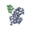
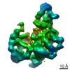
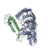
A: DNA polymerase epsilon subunit 2 B: DNA polymerase epsilon catalytic subunit A E: DNA polymerase epsilon subunit 2 F: DNA polymerase epsilon catalytic subunit A hetero molecules
DNApolymeraseepsilonsubunit2 / DNA polymerase II subunit 2 / DNA polymerase epsilon subunit B
Mass: 59600.887 Da / Num. of mol.: 2 Source method: isolated from a genetically manipulated source Source: (gene. exp.) Homo sapiens (human) / Gene: POLE2, DPE2 / Production host: Escherichia coli (E. coli) / References: UniProt: P56282, DNA-directed DNA polymerase
#2: Protein
DNApolymeraseepsiloncatalyticsubunitA / DNA polymerase II subunit A
Mass: 16517.137 Da / Num. of mol.: 2 / Fragment: UNP residues 2142-2286 Source method: isolated from a genetically manipulated source Source: (gene. exp.) Homo sapiens (human) / Gene: POLE, POLE1 / Production host: Escherichia coli (E. coli) / References: UniProt: Q07864, DNA-directed DNA polymerase
Protocol: SINGLE WAVELENGTH / Monochromatic (M) / Laue (L): M / Scattering type: x-ray
Radiation wavelength
Wavelength: 0.9792 Å / Relative weight: 1
Reflection
Resolution: 2.35→40 Å / Num. obs: 61804 / % possible obs: 98.6 % / Redundancy: 3.3 % / Biso Wilson estimate: 29 Å2 / Rmerge(I) obs: 0.072 / Net I/σ(I): 21.5
Reflection shell
Resolution: 2.35→2.39 Å / Redundancy: 3.1 % / Rmerge(I) obs: 0.481 / Mean I/σ(I) obs: 2.2 / Num. unique obs: 3044 / % possible all: 98.2
-
Processing
Software
Name
Version
Classification
CNS
1.1
refinement
HKL-2000
datareduction
HKL-2000
datascaling
PHENIX
phasing
Refinement
Method to determine structure: SAD / Resolution: 2.35→39.75 Å / Rfactor Rfree error: 0.01 / Data cutoff high absF: 3006919.42 / Data cutoff low absF: 0 / Cross valid method: THROUGHOUT / σ(F): 0
Rfactor
Num. reflection
% reflection
Selection details
Rfree
0.264
2958
5.1 %
RANDOM
Rwork
0.225
-
-
-
obs
0.225
58410
94.7 %
-
Solvent computation
Bsol: 38.3459 Å2 / ksol: 0.373505 e/Å3
Displacement parameters
Biso mean: 46 Å2
Baniso -1
Baniso -2
Baniso -3
1-
0.43 Å2
0 Å2
0 Å2
2-
-
-0.09 Å2
0 Å2
3-
-
-
-0.34 Å2
Refine analyze
Free
Obs
Luzzati coordinate error
0.39 Å
0.32 Å
Luzzati d res low
-
5 Å
Luzzati sigma a
0.52 Å
0.42 Å
Refinement step
Cycle: 1 / Resolution: 2.35→39.75 Å /
Protein
Nucleic acid
Ligand
Solvent
Total
Num. atoms
0
0
0
0
0
Refine LS restraints
Refine-ID
Type
Dev ideal
X-RAY DIFFRACTION
c_bond_d
0.009
X-RAY DIFFRACTION
c_bond_d_na
X-RAY DIFFRACTION
c_bond_d_prot
X-RAY DIFFRACTION
c_angle_d
X-RAY DIFFRACTION
c_angle_d_na
X-RAY DIFFRACTION
c_angle_d_prot
X-RAY DIFFRACTION
c_angle_deg
1.5
X-RAY DIFFRACTION
c_angle_deg_na
X-RAY DIFFRACTION
c_angle_deg_prot
X-RAY DIFFRACTION
c_dihedral_angle_d
23.9
X-RAY DIFFRACTION
c_dihedral_angle_d_na
X-RAY DIFFRACTION
c_dihedral_angle_d_prot
X-RAY DIFFRACTION
c_improper_angle_d
1
X-RAY DIFFRACTION
c_improper_angle_d_na
X-RAY DIFFRACTION
c_improper_angle_d_prot
X-RAY DIFFRACTION
c_mcbond_it
X-RAY DIFFRACTION
c_mcangle_it
X-RAY DIFFRACTION
c_scbond_it
X-RAY DIFFRACTION
c_scangle_it
LS refinement shell
Resolution: 2.35→2.5 Å / Rfactor Rfree error: 0.017 / Total num. of bins used: 6
Rfactor
Num. reflection
% reflection
Rfree
0.374
464
5.1 %
Rwork
0.329
8677
-
obs
-
-
90.2 %
Xplor file
Refine-ID
Serial no
Param file
Topol file
X-RAY DIFFRACTION
1
CNS_TOPPAR/protein_rep.param
CNS_TOPPAR/protein.top
X-RAY DIFFRACTION
2
CNS_TOPPAR/water_rep.param
CNS_TOPPAR/water.top
X-RAY DIFFRACTION
3
CNS_TOPPAR/ion.param
CNS_TOPPAR/iom.top
+
About Yorodumi
-
News
-
Feb 9, 2022. New format data for meta-information of EMDB entries
New format data for meta-information of EMDB entries
Version 3 of the EMDB header file is now the official format.
The previous official version 1.9 will be removed from the archive.
In the structure databanks used in Yorodumi, some data are registered as the other names, "COVID-19 virus" and "2019-nCoV". Here are the details of the virus and the list of structure data.
Jan 31, 2019. EMDB accession codes are about to change! (news from PDBe EMDB page)
EMDB accession codes are about to change! (news from PDBe EMDB page)
The allocation of 4 digits for EMDB accession codes will soon come to an end. Whilst these codes will remain in use, new EMDB accession codes will include an additional digit and will expand incrementally as the available range of codes is exhausted. The current 4-digit format prefixed with “EMD-” (i.e. EMD-XXXX) will advance to a 5-digit format (i.e. EMD-XXXXX), and so on. It is currently estimated that the 4-digit codes will be depleted around Spring 2019, at which point the 5-digit format will come into force.
The EM Navigator/Yorodumi systems omit the EMD- prefix.
Related info.:Q: What is EMD? / ID/Accession-code notation in Yorodumi/EM Navigator
Yorodumi is a browser for structure data from EMDB, PDB, SASBDB, etc.
This page is also the successor to EM Navigator detail page, and also detail information page/front-end page for Omokage search.
The word "yorodu" (or yorozu) is an old Japanese word meaning "ten thousand". "mi" (miru) is to see.
Related info.:EMDB / PDB / SASBDB / Comparison of 3 databanks / Yorodumi Search / Aug 31, 2016. New EM Navigator & Yorodumi / Yorodumi Papers / Jmol/JSmol / Function and homology information / Changes in new EM Navigator and Yorodumi
 Movie
Movie Controller

 Yorodumi
 Open data
 Basic information
 Components
 Keywords
 Function and homology information
 Homo sapiens (human)
 X-RAY DIFFRACTION /
  Authors
 United States, 3items
  Citation
 Structure visualization
 Downloads & links
 Other downloads

 PDBj
PDBj


 Assembly
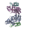


 Mass: 96.063 Da / Num. of mol.: 2 / Source method: obtained synthetically / Formula: SO4
 Mass: 65.409 Da / Num. of mol.: 4 / Source method: obtained synthetically / Formula: Zn
 Mass: 18.015 Da / Num. of mol.: 212 / Source method: isolated from a natural source / Formula: H2O
 Sample preparation
 Processing