 Movie
Movie Controller

[English] 日本語
 Yorodumi
- PDB-3e1h: Crystal structure of a type III polyketide synthase PKSIIINc from... -
+ Open data

- Basic information
| Entry | Database: PDB / ID: 3e1h | ||||||
|---|---|---|---|---|---|---|---|
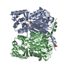
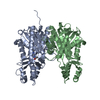
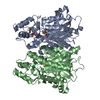
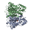
| Title | Crystal structure of a type III polyketide synthase PKSIIINc from Neurospora crassa | ||||||
 Components Components | Putative uncharacterized protein | ||||||
 Keywords Keywords | TRANSFERASE / Resorcinolic lipid synthase / Type III PKS / Acyltransferase | ||||||
| Function / homology |  Function and homology information Function and homology informationacyltransferase activity, transferring groups other than amino-acyl groups Similarity search - Function | ||||||
| Biological species |  Neurospora crassa (fungus) Neurospora crassa (fungus) | ||||||
| Method |  X-RAY DIFFRACTION / MOLECULAR REPLACEMENT / Resolution: 2.58 Å X-RAY DIFFRACTION / MOLECULAR REPLACEMENT / Resolution: 2.58 Å | ||||||
 Authors Authors | Goyal, A. / Rahman, A. / Sankaranarayanan, R. | ||||||
 Citation Citation | Journal: J.Struct.Biol. / Year: 2008 Title: Structural insights into biosynthesis of resorcinolic lipids by a type III polyketide synthase in Neurospora crassa Authors: Goyal, A. / Saxena, P. / Rahman, A. / Singh, P.K. / Kasbekar, D.P. / Gokhale, R.S. / Sankaranarayanan, R. | ||||||
| History |
|
- Structure visualization
| Structure viewer | Molecule: MolmilJmol/JSmol |
|---|
- Downloads & links
-Download
| PDBx/mmCIF format | 3e1h.cif.gz | 164.4 KB | Display | PDBx/mmCIF format |
|---|---|---|---|---|
| PDB format | pdb3e1h.ent.gz | 126.8 KB | Display | PDB format |
| PDBx/mmJSON format | 3e1h.json.gz | Tree view | PDBx/mmJSON format | |
| Others |  Other downloads Other downloads |
-Validation report
| Arichive directory | https://data.pdbj.org/pub/pdb/validation_reports/e1/3e1hftp://data.pdbj.org/pub/pdb/validation_reports/e1/3e1h | HTTPS FTP |
|---|
-Related structure data


-Links
 PDBj
PDBj

- Assembly
| Deposited unit | 
| ||||||||
|---|---|---|---|---|---|---|---|---|---|
| 1 |
| ||||||||
| Unit cell |
|
-Components
| #1: Protein | Mass: 49592.883 Da / Num. of mol.: 2 Source method: isolated from a genetically manipulated source Source: (gene. exp.) Neurospora crassa (fungus) / Strain: 74-OR23-1A (FGSC 987) / Plasmid: pET28c / Production host:  #2: Water | ChemComp-HOH / |  Mass: 18.015 Da / Num. of mol.: 413 / Source method: isolated from a natural source / Formula: H2O Mass: 18.015 Da / Num. of mol.: 413 / Source method: isolated from a natural source / Formula: H2O |
|---|
-Experimental details
-Experiment
| Experiment | Method: X-RAY DIFFRACTION / Number of used crystals: 1 |
|---|
- Sample preparation
| Crystal | Density Matthews: 1.96 Å3/Da / Density % sol: 37.17 % |
|---|---|
| Crystal grow | Temperature: 295 K / Method: vapor diffusion, hanging drop / pH: 6.8 Details: PEG 8000, Isopropanol, Sodium acetate, HEPES, pH 6.8, VAPOR DIFFUSION, HANGING DROP, temperature 295K |
-Data collection
| Diffraction | Mean temperature: 100 K |
|---|---|
| Diffraction source | Source: ROTATING ANODE / Type: RIGAKU RUH3R / Wavelength: 1.5418 Å |
| Detector | Type: MAR scanner 345 mm plate / Detector: IMAGE PLATE / Date: Jul 27, 2007 / Details: Osmic mirrors |
| Radiation | Protocol: SINGLE WAVELENGTH / Monochromatic (M) / Laue (L): M / Scattering type: x-ray |
| Radiation wavelength | Wavelength: 1.5418 Å / Relative weight: 1 |
| Reflection | Resolution: 2.58→25 Å / Num. all: 24396 / Num. obs: 24396 / % possible obs: 97.6 % / Redundancy: 4.4 % / Rmerge(I) obs: 0.091 |
| Reflection shell | Resolution: 2.58→2.67 Å / Redundancy: 3.5 % / Rmerge(I) obs: 0.387 / Mean I/σ(I) obs: 2.24 / Num. unique all: 2150 / % possible all: 88.2 |
- Processing
| Software |
| ||||||||||||||||||||||||||||||||||||||||||||||||||||||||||||||||||||||||||||||||||||||||||
|---|---|---|---|---|---|---|---|---|---|---|---|---|---|---|---|---|---|---|---|---|---|---|---|---|---|---|---|---|---|---|---|---|---|---|---|---|---|---|---|---|---|---|---|---|---|---|---|---|---|---|---|---|---|---|---|---|---|---|---|---|---|---|---|---|---|---|---|---|---|---|---|---|---|---|---|---|---|---|---|---|---|---|---|---|---|---|---|---|---|---|---|
| Refinement | Method to determine structure: MOLECULAR REPLACEMENT Starting model: PDB ENTRY 1CGZ and 1TED Resolution: 2.58→25 Å / Cor.coef. Fo:Fc: 0.922 / Cor.coef. Fo:Fc free: 0.896 / SU B: 11.38 / SU ML: 0.251 / Cross valid method: THROUGHOUT / ESU R Free: 0.357 / Stereochemistry target values: MAXIMUM LIKELIHOOD / Details: HYDROGENS HAVE BEEN ADDED IN THE RIDING POSITIONS
| ||||||||||||||||||||||||||||||||||||||||||||||||||||||||||||||||||||||||||||||||||||||||||
| Solvent computation | Ion probe radii: 0.8 Å / Shrinkage radii: 0.8 Å / VDW probe radii: 1.4 Å / Solvent model: MASK | ||||||||||||||||||||||||||||||||||||||||||||||||||||||||||||||||||||||||||||||||||||||||||
| Displacement parameters | Biso mean: 32.068 Å2
| ||||||||||||||||||||||||||||||||||||||||||||||||||||||||||||||||||||||||||||||||||||||||||
| Refinement step | Cycle: LAST / Resolution: 2.58→25 Å
| ||||||||||||||||||||||||||||||||||||||||||||||||||||||||||||||||||||||||||||||||||||||||||
| Refine LS restraints |
| ||||||||||||||||||||||||||||||||||||||||||||||||||||||||||||||||||||||||||||||||||||||||||
| LS refinement shell | Resolution: 2.591→2.658 Å / Total num. of bins used: 20
|
