 Movie
Movie Controller

[English] 日本語
 Yorodumi
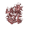
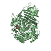
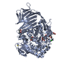
- PDB-1ted: Crystal structure of a type III polyketide synthase PKS18 from My... -
+ Open data

- Basic information
| Entry | Database: PDB / ID: 1ted | ||||||
|---|---|---|---|---|---|---|---|
| Title | Crystal structure of a type III polyketide synthase PKS18 from Mycobacterium tuberculosis | ||||||
 Components Components | pks18 | ||||||
 Keywords Keywords | TRANSFERASE / thiolase fold / substrate binding tunnel | ||||||
| Function / homology |  Function and homology information Function and homology informationchalcone biosynthetic process / polyketide biosynthetic process / acyltransferase activity, transferring groups other than amino-acyl groups / Transferases; Acyltransferases; Transferring groups other than aminoacyl groups / fatty acid biosynthetic process Similarity search - Function | ||||||
| Biological species |   Mycobacterium tuberculosis (bacteria) Mycobacterium tuberculosis (bacteria) | ||||||
| Method |  X-RAY DIFFRACTION / MOLECULAR REPLACEMENT / Resolution: 2.25 Å X-RAY DIFFRACTION / MOLECULAR REPLACEMENT / Resolution: 2.25 Å | ||||||
 Authors Authors | Sankaranarayanan, R. / Shanmugam, V.M. / Rukmini, R. | ||||||
 Citation Citation | Journal: Nat.Struct.Mol.Biol. / Year: 2004 Title: A novel tunnel in mycobacterial type III polyketide synthase reveals the structural basis for generating diverse metabolites Authors: Sankaranarayanan, R. / Saxena, P. / Marathe, U.B. / Gokhale, R.S. / Shanmugam, V.M. / Rukmini, R. #1: Journal: Acta Crystallogr.,Sect.D / Year: 2004 Title: Crystallization and preliminary X-ray crystallographic investigations of an unusual type III polyketide synthase PKS18 from Mycobacterium tuberculosis Authors: Rukmini, R. / Shanmugam, V.M. / Saxena, P. / Gokhale, R.S. / Sankaranarayanan, R. | ||||||
| History |
|
- Structure visualization
| Structure viewer | Molecule: MolmilJmol/JSmol |
|---|
- Downloads & links
-Download
| PDBx/mmCIF format | 1ted.cif.gz | 302.8 KB | Display | PDBx/mmCIF format |
|---|---|---|---|---|
| PDB format | pdb1ted.ent.gz | 242.5 KB | Display | PDB format |
| PDBx/mmJSON format | 1ted.json.gz | Tree view | PDBx/mmJSON format | |
| Others |  Other downloads Other downloads |
-Validation report
| Arichive directory | https://data.pdbj.org/pub/pdb/validation_reports/te/1tedftp://data.pdbj.org/pub/pdb/validation_reports/te/1ted | HTTPS FTP |
|---|
-Related structure data
| Related structure data |  1teeC  1cgzS C: citing same article ( S: Starting model for refinement |
|---|---|
| Similar structure data |


-Links
 PDBj
PDBj


- Assembly
| Deposited unit | 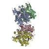
| ||||||||
|---|---|---|---|---|---|---|---|---|---|
| 1 | 
| ||||||||
| 2 | 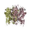
| ||||||||
| Unit cell |
| ||||||||
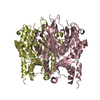
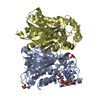
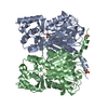
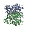
| Details | The biological assembly is a homodimer. The file contains two of them, AB and CD. |
-Components
| #1: Protein | Mass: 42073.316 Da / Num. of mol.: 4 Source method: isolated from a genetically manipulated source Source: (gene. exp.) Mycobacterium tuberculosis (bacteria) / Strain: H37Rv / Gene: pks18 / Plasmid: pET21C / Species (production host): Escherichia coli / Production host: References: UniProt: Q7D8I1, UniProt: P9WPF1*PLUS, chalcone synthase #2: Chemical | ChemComp-MYR /   Mass: 228.371 Da / Num. of mol.: 4 / Source method: obtained synthetically / Formula: C14H28O2 Mass: 228.371 Da / Num. of mol.: 4 / Source method: obtained synthetically / Formula: C14H28O2#3: Water | ChemComp-HOH / |  Mass: 18.015 Da / Num. of mol.: 1106 / Source method: isolated from a natural source / Formula: H2O Mass: 18.015 Da / Num. of mol.: 1106 / Source method: isolated from a natural source / Formula: H2O |
|---|
-Experimental details
-Experiment
| Experiment | Method: X-RAY DIFFRACTION / Number of used crystals: 1 |
|---|
- Sample preparation
| Crystal | Density Matthews: 2.5 Å3/Da / Density % sol: 50.4 % |
|---|---|
| Crystal grow | Temperature: 277 K / Method: vapor diffusion, hanging drop / pH: 7.7 Details: PEG 8000, HEPES, ethylene glycol, pH 7.7, VAPOR DIFFUSION, HANGING DROP, temperature 277K |
-Data collection
| Diffraction | Mean temperature: 100 K |
|---|---|
| Diffraction source | Source: ROTATING ANODE / Type: RIGAKU RU300 / Wavelength: 1.5418 Å |
| Detector | Type: MARRESEARCH / Detector: IMAGE PLATE / Date: Sep 26, 2003 |
| Radiation | Protocol: SINGLE WAVELENGTH / Monochromatic (M) / Laue (L): M / Scattering type: x-ray |
| Radiation wavelength | Wavelength: 1.5418 Å / Relative weight: 1 |
| Reflection | Resolution: 2.25→25 Å / Num. all: 80874 / Num. obs: 76912 / % possible obs: 95.1 % / Observed criterion σ(F): 0 / Observed criterion σ(I): 0 / Redundancy: 1.74 % / Biso Wilson estimate: 35.5 Å2 / Rmerge(I) obs: 0.07 / Net I/σ(I): 9.5 |
| Reflection shell | Resolution: 2.25→2.36 Å / Redundancy: 1.7 % / Rmerge(I) obs: 0.307 / Mean I/σ(I) obs: 2 / Num. unique all: 7360 / % possible all: 91.2 |
- Processing
| Software |
| ||||||||||||||||||||||||||||||||||||
|---|---|---|---|---|---|---|---|---|---|---|---|---|---|---|---|---|---|---|---|---|---|---|---|---|---|---|---|---|---|---|---|---|---|---|---|---|---|
| Refinement | Method to determine structure: MOLECULAR REPLACEMENT Starting model: PDB ENTRY 1CGZ Resolution: 2.25→24.87 Å / Rfactor Rfree error: 0.004 / Data cutoff high absF: 1677848.92 / Data cutoff low absF: 0 / Isotropic thermal model: RESTRAINED / Cross valid method: THROUGHOUT / σ(F): 0 / Stereochemistry target values: Engh & Huber
| ||||||||||||||||||||||||||||||||||||
| Solvent computation | Solvent model: FLAT MODEL / Bsol: 57.9511 Å2 / ksol: 0.328374 e/Å3 | ||||||||||||||||||||||||||||||||||||
| Displacement parameters | Biso mean: 36.7 Å2
| ||||||||||||||||||||||||||||||||||||
| Refine analyze |
| ||||||||||||||||||||||||||||||||||||
| Refinement step | Cycle: LAST / Resolution: 2.25→24.87 Å
| ||||||||||||||||||||||||||||||||||||
| Refine LS restraints |
| ||||||||||||||||||||||||||||||||||||
| LS refinement shell | Resolution: 2.25→2.33 Å / Rfactor Rfree error: 0.017 / Total num. of bins used: 10
| ||||||||||||||||||||||||||||||||||||
| Xplor file |
|
