 Movie
Movie Controller


+ Open data

- Basic information
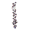
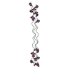
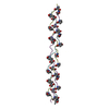
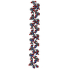
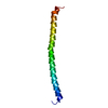
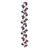
| Entry | Database: PDB / ID: 1bkv | ||||||
|---|---|---|---|---|---|---|---|
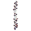
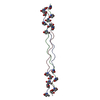
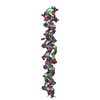
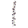
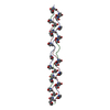
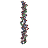
| Title | COLLAGEN | ||||||
 Components Components | T3-785 | ||||||
 Keywords Keywords | STRUCTURAL PROTEIN / COLLAGEN / HYDROXYPROLINE / HYDROGEN BONDING / TRIPLE HELIX / TYPE III COLLAGEN | ||||||
| Function / homology | ACETIC ACID Function and homology information Function and homology information | ||||||
| Method |  X-RAY DIFFRACTION / MOLECULAR REPLACEMENT / Resolution: 2 Å X-RAY DIFFRACTION / MOLECULAR REPLACEMENT / Resolution: 2 Å | ||||||
 Authors Authors | Kramer, R.Z. / Bella, J. / Mayville, P. / Brodsky, B. / Berman, H.M. | ||||||
 Citation Citation | Journal: Nat.Struct.Biol. / Year: 1999 Title: Sequence dependent conformational variations of collagen triple-helical structure. Authors: Kramer, R.Z. / Bella, J. / Mayville, P. / Brodsky, B. / Berman, H.M. | ||||||
| History |
|
- Structure visualization
| Structure viewer | Molecule: MolmilJmol/JSmol |
|---|
- Downloads & links
-Download
| PDBx/mmCIF format | 1bkv.cif.gz | 27.2 KB | Display | PDBx/mmCIF format |
|---|---|---|---|---|
| PDB format | pdb1bkv.ent.gz | 20.6 KB | Display | PDB format |
| PDBx/mmJSON format | 1bkv.json.gz | Tree view | PDBx/mmJSON format | |
| Others |  Other downloads Other downloads |
-Validation report
| Arichive directory | https://data.pdbj.org/pub/pdb/validation_reports/bk/1bkvftp://data.pdbj.org/pub/pdb/validation_reports/bk/1bkv | HTTPS FTP |
|---|
-Related structure data
| Similar structure data |
|---|

-Links
 PDBj
PDBj

- Assembly
| Deposited unit | 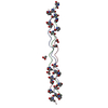
| ||||||||||||||||||||||||
|---|---|---|---|---|---|---|---|---|---|---|---|---|---|---|---|---|---|---|---|---|---|---|---|---|---|
| 1 |
| ||||||||||||||||||||||||
| Unit cell |
| ||||||||||||||||||||||||
| Components on special symmetry positions |
|
-Components
| #1: Protein/peptide | Mass: 2686.908 Da / Num. of mol.: 3 Source method: isolated from a genetically manipulated source Details: MODEL PEPTIDE CONTAINING BIOLOGICAL SEQUENCE WAS CHEMICALLY SYNTHESIZED #2: Chemical | ChemComp-ACY /   Mass: 60.052 Da / Num. of mol.: 5 / Source method: obtained synthetically / Formula: C2H4O2 Mass: 60.052 Da / Num. of mol.: 5 / Source method: obtained synthetically / Formula: C2H4O2#3: Water | ChemComp-HOH / |  Mass: 18.015 Da / Num. of mol.: 111 / Source method: isolated from a natural source / Formula: H2O Mass: 18.015 Da / Num. of mol.: 111 / Source method: isolated from a natural source / Formula: H2OCompound details | HYDROGEN BONDS BETWEEN PEPTIDE CHAINS FOLLOW THE RICH AND CRICK MODEL II FOR COLLAGEN. | Nonpolymer details | TWO ACETIC ACID MOLECULES SIT ON A CRYSTALLOGRAPHIC TWO FOLD. AS A RESULT THE ASYMMETRIC UNIT FOR ...TWO ACETIC ACID MOLECULES SIT ON A CRYSTALLOG | |
|---|
-Experimental details
-Experiment
| Experiment | Method: X-RAY DIFFRACTION / Number of used crystals: 1 |
|---|
- Sample preparation
| Crystal | Density Matthews: 2.1 Å3/Da / Density % sol: 43.5 % | ||||||||||||||||||||||||||||||||||||||||||||||||||||||
|---|---|---|---|---|---|---|---|---|---|---|---|---|---|---|---|---|---|---|---|---|---|---|---|---|---|---|---|---|---|---|---|---|---|---|---|---|---|---|---|---|---|---|---|---|---|---|---|---|---|---|---|---|---|---|---|
| Crystal grow | pH: 8.5 / Details: pH 8.50 | ||||||||||||||||||||||||||||||||||||||||||||||||||||||
| Crystal grow | *PLUS Temperature: 4 ℃ / Method: vapor diffusion, hanging drop | ||||||||||||||||||||||||||||||||||||||||||||||||||||||
| Components of the solutions | *PLUS
|
-Data collection
| Diffraction | Mean temperature: 108 K |
|---|---|
| Diffraction source | Source: ROTATING ANODE / Type: RIGAKU RUH2R / Wavelength: 1.5418 |
| Detector | Type: RIGAKU RAXIS IV / Detector: IMAGE PLATE / Date: Dec 1, 1996 / Details: MIRRORS |
| Radiation | Protocol: SINGLE WAVELENGTH / Monochromatic (M) / Laue (L): M / Scattering type: x-ray |
| Radiation wavelength | Wavelength: 1.5418 Å / Relative weight: 1 |
| Reflection | Resolution: 2→50 Å / Num. obs: 4853 / % possible obs: 95.9 % / Observed criterion σ(I): 0 / Rmerge(I) obs: 0.053 |
| Reflection shell | Resolution: 2→2.06 Å / Rmerge(I) obs: 0.133 / % possible all: 93 |
| Reflection | *PLUS % possible obs: 96 % / Rmerge(I) obs: 0.058 |
| Reflection shell | *PLUS % possible obs: 85 % |
- Processing
| Software |
| ||||||||||||||||||||||||||||||||||||||||||||||||||||||||||||
|---|---|---|---|---|---|---|---|---|---|---|---|---|---|---|---|---|---|---|---|---|---|---|---|---|---|---|---|---|---|---|---|---|---|---|---|---|---|---|---|---|---|---|---|---|---|---|---|---|---|---|---|---|---|---|---|---|---|---|---|---|---|
| Refinement | Method to determine structure: MOLECULAR REPLACEMENT Starting model: IDEALIZED 7-FOLD TRIPLE HELIX Resolution: 2→50 Å / Cross valid method: THROUGHOUT / σ(F): 2 Details: ADDITIONAL PARAMETERS USED FOR HYDROXYPROLINE. THREE EXTREMELY STRONG REFLECTIONS (-3 1 1, 4 0 2, AND 5 1 0) WERE UNDERESTIMATED AND HENCE WERE EXCLUDED FROM REFINEMENT.
| ||||||||||||||||||||||||||||||||||||||||||||||||||||||||||||
| Displacement parameters | Biso mean: 36.4 Å2
| ||||||||||||||||||||||||||||||||||||||||||||||||||||||||||||
| Refinement step | Cycle: LAST / Resolution: 2→50 Å
| ||||||||||||||||||||||||||||||||||||||||||||||||||||||||||||
| Refine LS restraints |
| ||||||||||||||||||||||||||||||||||||||||||||||||||||||||||||
| LS refinement shell | Resolution: 2→2.07 Å / Total num. of bins used: 10
| ||||||||||||||||||||||||||||||||||||||||||||||||||||||||||||
| Xplor file | Serial no: 1 / Param file: PROTEIN_REP.PARAM / Topol file: TOPHCSDX.PRO | ||||||||||||||||||||||||||||||||||||||||||||||||||||||||||||
| Software | *PLUS Name: CNS / Version: 0.3 / Classification: refinement | ||||||||||||||||||||||||||||||||||||||||||||||||||||||||||||
| Refine LS restraints | *PLUS
|
