 Movie
Movie Controller


+ Open data

- Basic information
| Entry | Database: PDB / ID: 1b6g | ||||||
|---|---|---|---|---|---|---|---|
| Title | HALOALKANE DEHALOGENASE AT PH 5.0 CONTAINING CHLORIDE | ||||||
 Components Components | HALOALKANE DEHALOGENASE | ||||||
 Keywords Keywords | HYDROLASE / HALOALKANE DEHALOGENASE / ALPHA/BETA-HYDROLASE | ||||||
| Function / homology |  Function and homology information Function and homology information1,2-dichloroethane catabolic process / haloalkane dehalogenase / haloalkane dehalogenase activity / epoxide hydrolase activity / response to toxic substance Similarity search - Function | ||||||
| Biological species |  Xanthobacter autotrophicus (bacteria) Xanthobacter autotrophicus (bacteria) | ||||||
| Method |  X-RAY DIFFRACTION / SYNCHROTRON / MOLECULAR REPLACEMENT / Resolution: 1.15 Å X-RAY DIFFRACTION / SYNCHROTRON / MOLECULAR REPLACEMENT / Resolution: 1.15 Å | ||||||
 Authors Authors | Ridder, I.S. / Rozeboom, H.J. / Dijkstra, B.W. | ||||||
 Citation Citation | Journal: Acta Crystallogr.,Sect.D / Year: 1999 Title: Haloalkane dehalogenase from Xanthobacter autotrophicus GJ10 refined at 1.15 A resolution. Authors: Ridder, I.S. / Rozeboom, H.J. / Dijkstra, B.W. #1: Journal: Biochemistry / Year: 1998Title: Kinetic Analysis and X-Ray Structure of Haloalkane Dehalogenase with a Modified Halide-Binding Site Authors: Krooshof, G.H. / Ridder, I.S. / Tepper, A.W. / Vos, G.J. / Rozeboom, H.J. / Kalk, K.H. / Dijkstra, B.W. / Janssen, D.B. #2: Journal: Nature / Year: 1993Title: Crystallographic Analysis of the Catalytic Mechanism of Haloalkane Dehalogenase Authors: Verschueren, K.H. / Seljee, F. / Rozeboom, H.J. / Kalk, K.H. / Dijkstra, B.W. #3: Journal: J.Mol.Biol. / Year: 1993Title: Refined X-Ray Structures of Haloalkane Dehalogenase at Ph 6.2 And Ph 8.2 And Implications for the Reaction Mechanism Authors: Verschueren, K.H. / Franken, S.M. / Rozeboom, H.J. / Kalk, K.H. / Dijkstra, B.W. #4: Journal: Embo J. / Year: 1991Title: Crystal Structure of Haloalkane Dehalogenase: An Enzyme to Detoxify Halogenated Alkanes Authors: Franken, S.M. / Rozeboom, H.J. / Kalk, K.H. / Dijkstra, B.W. #5: Journal: J.Mol.Biol. / Year: 1988Title: Crystallization of Haloalkane Dehalogenase from Xanthobacter Autotrophicus Gj10 Authors: Rozeboom, H.J. / Kingma, J. / Janssen, D.B. / Dijkstra, B.W. | ||||||
| History |
|
- Structure visualization
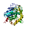
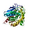
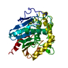
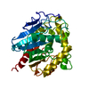
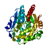
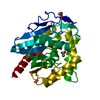
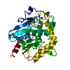
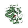
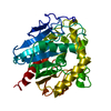
| Structure viewer | Molecule: MolmilJmol/JSmol |
|---|
- Downloads & links
-Download
| PDBx/mmCIF format | 1b6g.cif.gz | 171.4 KB | Display | PDBx/mmCIF format |
|---|---|---|---|---|
| PDB format | pdb1b6g.ent.gz | 135.7 KB | Display | PDB format |
| PDBx/mmJSON format | 1b6g.json.gz | Tree view | PDBx/mmJSON format | |
| Others |  Other downloads Other downloads |
-Validation report
| Summary document | 1b6g_validation.pdf.gz | 389.6 KB | Display | wwPDB validaton report |
|---|---|---|---|---|
| Full document | 1b6g_full_validation.pdf.gz | 395.3 KB | Display | |
| Data in XML | 1b6g_validation.xml.gz | 9.5 KB | Display | |
| Data in CIF | 1b6g_validation.cif.gz | 17.1 KB | Display | |
| Arichive directory | https://data.pdbj.org/pub/pdb/validation_reports/b6/1b6gftp://data.pdbj.org/pub/pdb/validation_reports/b6/1b6g | HTTPS FTP |
-Related structure data
| Related structure data | 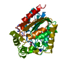 1be0S S: Starting model for refinement |
|---|---|
| Similar structure data |

-Links
 PDBj
PDBj


- Assembly
| Deposited unit | 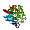
| ||||||||
|---|---|---|---|---|---|---|---|---|---|
| 1 |
| ||||||||
| Unit cell |
| ||||||||
| Components on special symmetry positions |
|
-Components
| #1: Protein | Mass: 35574.152 Da / Num. of mol.: 1 Source method: isolated from a genetically manipulated source Source: (gene. exp.) Xanthobacter autotrophicus (bacteria) / Strain: GJ10 / Production host: | ||||||
|---|---|---|---|---|---|---|---|
| #2: Chemical | ChemComp-SO4 /   Mass: 96.063 Da / Num. of mol.: 1 / Source method: obtained synthetically / Formula: SO4 Mass: 96.063 Da / Num. of mol.: 1 / Source method: obtained synthetically / Formula: SO4 | ||||||
| #3: Chemical |   Mass: 35.453 Da / Num. of mol.: 2 / Source method: obtained synthetically / Formula: Cl Mass: 35.453 Da / Num. of mol.: 2 / Source method: obtained synthetically / Formula: Cl#4: Chemical | ChemComp-GOL /   Mass: 92.094 Da / Num. of mol.: 7 / Source method: obtained synthetically / Formula: C3H8O3 Mass: 92.094 Da / Num. of mol.: 7 / Source method: obtained synthetically / Formula: C3H8O3#5: Water | ChemComp-HOH / |  Mass: 18.015 Da / Num. of mol.: 601 / Source method: isolated from a natural source / Formula: H2O Mass: 18.015 Da / Num. of mol.: 601 / Source method: isolated from a natural source / Formula: H2OHas protein modification | Y | |
-Experimental details
-Experiment
| Experiment | Method: X-RAY DIFFRACTION / Number of used crystals: 1 |
|---|
- Sample preparation
| Crystal | Density Matthews: 1.89 Å3/Da / Density % sol: 35 % | ||||||||||||||||||||||||||||||||||||
|---|---|---|---|---|---|---|---|---|---|---|---|---|---|---|---|---|---|---|---|---|---|---|---|---|---|---|---|---|---|---|---|---|---|---|---|---|---|
| Crystal grow | pH: 5 / Details: pH 5.0 | ||||||||||||||||||||||||||||||||||||
| Crystal grow | *PLUS Method: vapor diffusion, hanging drop | ||||||||||||||||||||||||||||||||||||
| Components of the solutions | *PLUS
|
-Data collection
| Diffraction | Mean temperature: 100 K |
|---|---|
| Diffraction source | Source: SYNCHROTRON / Site: ESRF  / Beamline: ID14-3 / Wavelength: 0.9475 / Beamline: ID14-3 / Wavelength: 0.9475 |
| Detector | Type: MARRESEARCH / Detector: CCD / Date: Apr 6, 1998 |
| Radiation | Monochromatic (M) / Laue (L): M / Scattering type: x-ray |
| Radiation wavelength | Wavelength: 0.9475 Å / Relative weight: 1 |
| Reflection | Resolution: 1.152→30 Å / Num. obs: 94837 / % possible obs: 97.6 % / Redundancy: 3.67 % / Rmerge(I) obs: 0.039 / Net I/σ(I): 30.8 |
| Reflection shell | Resolution: 1.15→1.17 Å / Redundancy: 3.52 % / Rmerge(I) obs: 0.316 / Mean I/σ(I) obs: 2.7 / % possible all: 92.5 |
| Reflection | *PLUS Num. measured all: 347849 |
| Reflection shell | *PLUS % possible obs: 92.5 % |
- Processing
| Software |
| |||||||||||||||||||||||||||||||||
|---|---|---|---|---|---|---|---|---|---|---|---|---|---|---|---|---|---|---|---|---|---|---|---|---|---|---|---|---|---|---|---|---|---|---|
| Refinement | Method to determine structure: MOLECULAR REPLACEMENT Starting model: 1BE0 Resolution: 1.15→15 Å / Num. parameters: 29678 / Num. restraintsaints: 36636 / Cross valid method: FREE R / σ(F): 0 / Stereochemistry target values: ENGH AND HUBER Details: ANISOTROPIC REFINEMENT REDUCED RWORK (NO CUTOFF) FROM 16.4% TO 13.4% AND RFREE FROM 19.6% TO 17.3%. THE FOLLOWING ARE APPARENT CLOSE CONTACTS BUT ACTUALLY INVOLVE DIFFERENT ALTERNATE ...Details: ANISOTROPIC REFINEMENT REDUCED RWORK (NO CUTOFF) FROM 16.4% TO 13.4% AND RFREE FROM 19.6% TO 17.3%. THE FOLLOWING ARE APPARENT CLOSE CONTACTS BUT ACTUALLY INVOLVE DIFFERENT ALTERNATE CONFORMATIONS: CLOSE CONTACT C3 GOL 1204 - O HOH 2608 0.604 CLOSE CONTACT CD AGLU 280 - O HOH 2586 0.938 CLOSE CONTACT CE BLYS 192 - O HOH 2549 0.662 CLOSE CONTACT NH1BARG 300 - O HOH 2607 1.006 CLOSE CONTACT NZ BLYS 192 - O HOH 2549 0.874 CLOSE CONTACT O HOH 2558 - O HOH 2565 1.168 CLOSE CONTACT O HOH 2565 - O HOH 2558 1.168 CLOSE CONTACT OD1BASP 137 - O HOH 2585 0.681 CLOSE CONTACT OD2BASP 137 - O HOH 2584 0.868 CLOSE CONTACT OE1AGLU 280 - O HOH 2586 0.955 CLOSE CONTACT OE1BGLU 72 - O HOH 2591 1.117 CLOSE CONTACTCL CL 1999 - O HOH 2000 0.267
| |||||||||||||||||||||||||||||||||
| Solvent computation | Solvent model: MOEWS & KRETSINGER, J.MOL.BIOL.91(1973)201-2 | |||||||||||||||||||||||||||||||||
| Refine analyze | Num. disordered residues: 34 / Occupancy sum hydrogen: 2406.8 / Occupancy sum non hydrogen: 3064.8 | |||||||||||||||||||||||||||||||||
| Refinement step | Cycle: LAST / Resolution: 1.15→15 Å
| |||||||||||||||||||||||||||||||||
| Refine LS restraints |
|
