 Movie
Movie Controller

[English] 日本語
 Yorodumi
- PDB-2yxp: The Effect of Deuteration on Protein Structure A High Resolution ... -
+ Open data

- Basic information
| Entry | Database: PDB / ID: 2yxp | ||||||
|---|---|---|---|---|---|---|---|
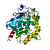
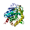
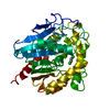
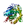
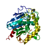
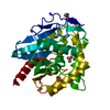
| Title | The Effect of Deuteration on Protein Structure A High Resolution Comparison of Hydrogenous and Perdeuterated Haloalkane Dehalogenase | ||||||
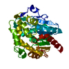
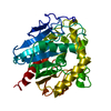
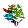
 Components Components | Haloalkane dehalogenase | ||||||
 Keywords Keywords | HYDROLASE / protein deuteration / haloalkane dehalogenase / high resolution structure / catalytic mechanism | ||||||
| Function / homology |  Function and homology information Function and homology information1,2-dichloroethane catabolic process / haloalkane dehalogenase / haloalkane dehalogenase activity / epoxide hydrolase activity / response to toxic substance Similarity search - Function | ||||||
| Biological species |  Xanthobacter autotrophicus (bacteria) Xanthobacter autotrophicus (bacteria) | ||||||
| Method |  X-RAY DIFFRACTION / rigid body refinement / Resolution: 1.53 Å X-RAY DIFFRACTION / rigid body refinement / Resolution: 1.53 Å | ||||||
 Authors Authors | Liu, X. / Hanson, L. / Langan, P. / Viola, R.E. | ||||||
 Citation Citation | Journal: Acta Crystallogr.,Sect.D / Year: 2007 Title: The effect of deuteration on protein structure: a high-resolution comparison of hydrogenous and perdeuterated haloalkane dehalogenase. Authors: Liu, X. / Hanson, B.L. / Langan, P. / Viola, R.E. | ||||||
| History |
|
- Structure visualization
| Structure viewer | Molecule: MolmilJmol/JSmol |
|---|
- Downloads & links
-Download
| PDBx/mmCIF format | 2yxp.cif.gz | 82.4 KB | Display | PDBx/mmCIF format |
|---|---|---|---|---|
| PDB format | pdb2yxp.ent.gz | 61.7 KB | Display | PDB format |
| PDBx/mmJSON format | 2yxp.json.gz | Tree view | PDBx/mmJSON format | |
| Others |  Other downloads Other downloads |
-Validation report
| Arichive directory | https://data.pdbj.org/pub/pdb/validation_reports/yx/2yxpftp://data.pdbj.org/pub/pdb/validation_reports/yx/2yxp | HTTPS FTP |
|---|
-Related structure data

-Links
 PDBj
PDBj


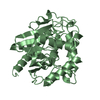
- Assembly
| Deposited unit | 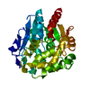
| ||||||||
|---|---|---|---|---|---|---|---|---|---|
| 1 |
| ||||||||
| Unit cell |
| ||||||||
| Components on special symmetry positions |
|
-Components
| #1: Protein | Mass: 35175.797 Da / Num. of mol.: 1 Source method: isolated from a genetically manipulated source Source: (gene. exp.) Xanthobacter autotrophicus (bacteria) / Strain: GJ10 / Gene: dhlA / Plasmid: pCH109 / Species (production host): Escherichia coli / Production host: |
|---|---|
| #2: Water | ChemComp-HOH /  Mass: 18.015 Da / Num. of mol.: 399 / Source method: isolated from a natural source / Formula: H2O Mass: 18.015 Da / Num. of mol.: 399 / Source method: isolated from a natural source / Formula: H2O |
-Experimental details
-Experiment
| Experiment | Method: X-RAY DIFFRACTION / Number of used crystals: 1 |
|---|
- Sample preparation
| Crystal | Density Matthews: 1.96 Å3/Da / Density % sol: 37.1 % |
|---|---|
| Crystal grow | Temperature: 293 K / Method: vapor diffusion, hanging drop / pH: 6 Details: 2.4M ammonium sulfate, 0.1M MES,, pH 6.0, VAPOR DIFFUSION, HANGING DROP, temperature 293K |
-Data collection
| Diffraction | Mean temperature: 100 K |
|---|---|
| Diffraction source | Source: ROTATING ANODE / Type: RIGAKU / Wavelength: 1.5418 Å |
| Detector | Type: RIGAKU RAXIS IV / Detector: IMAGE PLATE / Date: Sep 13, 2004 |
| Radiation | Monochromator: mirror / Protocol: SINGLE WAVELENGTH / Monochromatic (M) / Laue (L): M / Scattering type: x-ray |
| Radiation wavelength | Wavelength: 1.5418 Å / Relative weight: 1 |
| Reflection | Resolution: 1.53→37.62 Å / Num. obs: 36268 / % possible obs: 0.99 % / Observed criterion σ(F): 1.4 / Observed criterion σ(I): 1.4 / Redundancy: 6.12 % / Rsym value: 0.055 / Net I/σ(I): 17.5 |
| Reflection shell | Resolution: 1.53→1.59 Å / Redundancy: 2.05 % / Mean I/σ(I) obs: 3.1 / Num. unique all: 1996 / Rsym value: 0.257 / % possible all: 0.48 |
- Processing
| Software |
| |||||||||||||||||||||||||||||||||||||||||||||||||||||||||||||||||||||||||||||||||||||||||||||||||||||||||||||||||||||||||||||
|---|---|---|---|---|---|---|---|---|---|---|---|---|---|---|---|---|---|---|---|---|---|---|---|---|---|---|---|---|---|---|---|---|---|---|---|---|---|---|---|---|---|---|---|---|---|---|---|---|---|---|---|---|---|---|---|---|---|---|---|---|---|---|---|---|---|---|---|---|---|---|---|---|---|---|---|---|---|---|---|---|---|---|---|---|---|---|---|---|---|---|---|---|---|---|---|---|---|---|---|---|---|---|---|---|---|---|---|---|---|---|---|---|---|---|---|---|---|---|---|---|---|---|---|---|---|---|
| Refinement | Method to determine structure: rigid body refinement / Resolution: 1.53→37.62 Å / Cor.coef. Fo:Fc: 0.97 / Cor.coef. Fo:Fc free: 0.963 / SU B: 1.584 / SU ML: 0.057 / Cross valid method: THROUGHOUT / σ(I): 3.1 / ESU R: 0.092 / ESU R Free: 0.088 / Stereochemistry target values: MAXIMUM LIKELIHOOD
| |||||||||||||||||||||||||||||||||||||||||||||||||||||||||||||||||||||||||||||||||||||||||||||||||||||||||||||||||||||||||||||
| Solvent computation | Ion probe radii: 0.8 Å / Shrinkage radii: 0.8 Å / VDW probe radii: 1.4 Å / Solvent model: BABINET MODEL WITH MASK | |||||||||||||||||||||||||||||||||||||||||||||||||||||||||||||||||||||||||||||||||||||||||||||||||||||||||||||||||||||||||||||
| Displacement parameters | Biso mean: 18.281 Å2
| |||||||||||||||||||||||||||||||||||||||||||||||||||||||||||||||||||||||||||||||||||||||||||||||||||||||||||||||||||||||||||||
| Refinement step | Cycle: LAST / Resolution: 1.53→37.62 Å
| |||||||||||||||||||||||||||||||||||||||||||||||||||||||||||||||||||||||||||||||||||||||||||||||||||||||||||||||||||||||||||||
| Refine LS restraints |
| |||||||||||||||||||||||||||||||||||||||||||||||||||||||||||||||||||||||||||||||||||||||||||||||||||||||||||||||||||||||||||||
| LS refinement shell | Resolution: 1.53→1.57 Å / Total num. of bins used: 20
|
