 Movie
Movie Controller

[English] 日本語
 Yorodumi
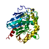
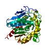
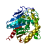
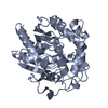
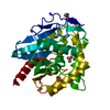
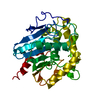
- PDB-1cij: HALOALKANE DEHALOGENASE SOAKED WITH HIGH CONCENTRATION OF BROMIDE -
+ Open data

- Basic information
| Entry | Database: PDB / ID: 1cij | ||||||
|---|---|---|---|---|---|---|---|
| Title | HALOALKANE DEHALOGENASE SOAKED WITH HIGH CONCENTRATION OF BROMIDE | ||||||
 Components Components | PROTEIN (HALOALKANE DEHALOGENASE) | ||||||
 Keywords Keywords | HYDROLASE / DEHALOGENASE / COLLISION COMPLEX / ALPHA/BETA-HYDROLASE | ||||||
| Function / homology |  Function and homology information Function and homology information1,2-dichloroethane catabolic process / haloalkane dehalogenase / haloalkane dehalogenase activity / epoxide hydrolase activity / response to toxic substance Similarity search - Function | ||||||
| Biological species |  Xanthobacter autotrophicus (bacteria) Xanthobacter autotrophicus (bacteria) | ||||||
| Method |  X-RAY DIFFRACTION / OTHER / Resolution: 2.3 Å X-RAY DIFFRACTION / OTHER / Resolution: 2.3 Å | ||||||
 Authors Authors | Ridder, I.S. / Rozeboom, H.J. / Kalk, K.H. / Dijkstra, B.W. | ||||||
 Citation Citation | Journal: Biochemistry / Year: 1999 Title: Crystallographic and kinetic evidence of a collision complex formed during halide import in haloalkane dehalogenase. Authors: Pikkemaat, M.G. / Ridder, I.S. / Rozeboom, H.J. / Kalk, K.H. / Dijkstra, B.W. / Janssen, D.B. #1: Journal: Nature / Year: 1993Title: Crystallographic analysis of the catalytic mechanism of haloalkane dehalogenase. Authors: Verschueren, K.H. / Seljee, F. / Rozeboom, H.J. / Kalk, K.H. / Dijkstra, B.W. #2: Journal: J.Mol.Biol. / Year: 1993Title: Refined X-ray structures of haloalkane dehalogenase at pH 6.2 and pH 8.2 and implications for the reaction mechanism. Authors: Verschueren, K.H. / Franken, S.M. / Rozeboom, H.J. / Kalk, K.H. / Dijkstra, B.W. #3: Journal: Embo J. / Year: 1991Title: Crystal structure of haloalkane dehalogenase: an enzyme to detoxify halogenated alkanes. Authors: Franken, S.M. / Rozeboom, H.J. / Kalk, K.H. / Dijkstra, B.W. #4: Journal: J.Mol.Biol. / Year: 1988 Title: Crystallization of haloalkane dehalogenase from Xanthobacter autotrophicus GJ10. Authors: Rozeboom, H.J. / Kingma, J. / Janssen, D.B. / Dijkstra, B.W. | ||||||
| History |
|
- Structure visualization
| Structure viewer | Molecule: MolmilJmol/JSmol |
|---|
- Downloads & links
-Download
| PDBx/mmCIF format | 1cij.cif.gz | 77.5 KB | Display | PDBx/mmCIF format |
|---|---|---|---|---|
| PDB format | pdb1cij.ent.gz | 57 KB | Display | PDB format |
| PDBx/mmJSON format | 1cij.json.gz | Tree view | PDBx/mmJSON format | |
| Others |  Other downloads Other downloads |
-Validation report
| Arichive directory | https://data.pdbj.org/pub/pdb/validation_reports/ci/1cijftp://data.pdbj.org/pub/pdb/validation_reports/ci/1cij | HTTPS FTP |
|---|
-Related structure data
| Related structure data | 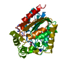 1be0S S: Starting model for refinement |
|---|---|
| Similar structure data |
-Links
 PDBj
PDBj


- Assembly
| Deposited unit | 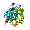
| ||||||||
|---|---|---|---|---|---|---|---|---|---|
| 1 |
| ||||||||
| Unit cell |
|
-Components
| #1: Protein | Mass: 35161.770 Da / Num. of mol.: 1 / Mutation: I2V Source method: isolated from a genetically manipulated source Details: BROMIDE ION / Source: (gene. exp.) Xanthobacter autotrophicus (bacteria) / Strain: GJ10 / Production host: | ||||
|---|---|---|---|---|---|
| #2: Chemical |   Mass: 79.904 Da / Num. of mol.: 3 / Source method: obtained synthetically / Formula: Br Mass: 79.904 Da / Num. of mol.: 3 / Source method: obtained synthetically / Formula: Br#3: Water | ChemComp-HOH / |  Mass: 18.015 Da / Num. of mol.: 160 / Source method: isolated from a natural source / Formula: H2O Mass: 18.015 Da / Num. of mol.: 160 / Source method: isolated from a natural source / Formula: H2OSequence details | DISCREPANC | |
-Experimental details
-Experiment
| Experiment | Method: X-RAY DIFFRACTION / Number of used crystals: 1 |
|---|
- Sample preparation
| Crystal | Density Matthews: 1.86 Å3/Da / Density % sol: 34 % | |||||||||||||||||||||||||
|---|---|---|---|---|---|---|---|---|---|---|---|---|---|---|---|---|---|---|---|---|---|---|---|---|---|---|
| Crystal grow | pH: 5.8 Details: 50 % AMMONIUM SULFATE 100 MM MES BUFFER, PH 5.9 , pH 5.8 | |||||||||||||||||||||||||
| Crystal | *PLUS | |||||||||||||||||||||||||
| Crystal grow | *PLUS Method: vapor diffusion, hanging drop / PH range low: 5.9 / PH range high: 5.7 | |||||||||||||||||||||||||
| Components of the solutions | *PLUS
|
-Data collection
| Diffraction | Mean temperature: 120 K |
|---|---|
| Diffraction source | Source: ROTATING ANODE / Type: ENRAF-NONIUS FR591 / Wavelength: 1.5418 |
| Detector | Type: MAC Science DIP-2030 / Detector: IMAGE PLATE / Date: Dec 24, 1997 / Details: DOUBLE MIRRORS (MAC-XOS) |
| Radiation | Monochromator: GRAPHITE / Protocol: SINGLE WAVELENGTH / Monochromatic (M) / Laue (L): M / Scattering type: x-ray |
| Radiation wavelength | Wavelength: 1.5418 Å / Relative weight: 1 |
| Reflection | Resolution: 2.3→50 Å / Num. obs: 12688 / % possible obs: 97.6 % / Observed criterion σ(I): 5 / Redundancy: 2.7 % / Biso Wilson estimate: 19.5 Å2 / Rsym value: 0.108 / Net I/σ(I): 8 |
| Reflection shell | Resolution: 2.3→2.34 Å / Redundancy: 2.5 % / Mean I/σ(I) obs: 2.1 / Rsym value: 0.363 / % possible all: 98.2 |
| Reflection | *PLUS Num. measured all: 34688 / Rmerge(I) obs: 0.108 |
| Reflection shell | *PLUS % possible obs: 98.2 % / Rmerge(I) obs: 0.363 |
- Processing
| Software |
| ||||||||||||||||||||||||||||||||||||||||||||||||||||||||||||||||||||||||||||||||
|---|---|---|---|---|---|---|---|---|---|---|---|---|---|---|---|---|---|---|---|---|---|---|---|---|---|---|---|---|---|---|---|---|---|---|---|---|---|---|---|---|---|---|---|---|---|---|---|---|---|---|---|---|---|---|---|---|---|---|---|---|---|---|---|---|---|---|---|---|---|---|---|---|---|---|---|---|---|---|---|---|---|
| Refinement | Method to determine structure: OTHER Starting model: PDB ENTRY 1BE0 Resolution: 2.3→20 Å / Data cutoff high absF: 100000 / Data cutoff low absF: 0.0001 / Isotropic thermal model: RESTRAINED Cross valid method: THROUGHOUT, EXCEPT LAST STEP IN WHICH ALL DATA (WORK+TEST SET) WERE USED σ(F): 0 Details: THE VALUES LISTED AS WORKING IN REMARK 3 APPLY TO THE LAST CYCLE OF THE REFINEMENT IN WHICH ALL DATA (WORK + FREE) WERE USED.
| ||||||||||||||||||||||||||||||||||||||||||||||||||||||||||||||||||||||||||||||||
| Displacement parameters | Biso mean: 16.2 Å2 | ||||||||||||||||||||||||||||||||||||||||||||||||||||||||||||||||||||||||||||||||
| Refine analyze | Luzzati sigma a obs: 0.3 Å | ||||||||||||||||||||||||||||||||||||||||||||||||||||||||||||||||||||||||||||||||
| Refinement step | Cycle: LAST / Resolution: 2.3→20 Å
| ||||||||||||||||||||||||||||||||||||||||||||||||||||||||||||||||||||||||||||||||
| Refine LS restraints |
| ||||||||||||||||||||||||||||||||||||||||||||||||||||||||||||||||||||||||||||||||
| LS refinement shell | Resolution: 2.3→2.4 Å / Total num. of bins used: 8
| ||||||||||||||||||||||||||||||||||||||||||||||||||||||||||||||||||||||||||||||||
| Xplor file |
| ||||||||||||||||||||||||||||||||||||||||||||||||||||||||||||||||||||||||||||||||
| Software | *PLUS Name: X-PLOR / Version: 3.843 / Classification: refinement | ||||||||||||||||||||||||||||||||||||||||||||||||||||||||||||||||||||||||||||||||
| Refinement | *PLUS Lowest resolution: 20 Å / σ(F): 0 / % reflection Rfree: 7.8 % | ||||||||||||||||||||||||||||||||||||||||||||||||||||||||||||||||||||||||||||||||
| Solvent computation | *PLUS | ||||||||||||||||||||||||||||||||||||||||||||||||||||||||||||||||||||||||||||||||
| Displacement parameters | *PLUS Biso mean: 16.2 Å2 | ||||||||||||||||||||||||||||||||||||||||||||||||||||||||||||||||||||||||||||||||
| Refine LS restraints | *PLUS
| ||||||||||||||||||||||||||||||||||||||||||||||||||||||||||||||||||||||||||||||||
| LS refinement shell | *PLUS Rfactor Rfree: 0.363 / % reflection Rfree: 8.8 % / Rfactor Rwork: 0.305 |
