 Movie
Movie Controller


+ Open data

- Basic information
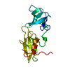
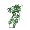
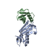
| Entry | Database: PDB / ID: 1ayi | ||||||
|---|---|---|---|---|---|---|---|
| Title | COLICIN E7 IMMUNITY PROTEIN IM7 | ||||||
 Components Components | COLICIN E IMMUNITY PROTEIN 7 | ||||||
 Keywords Keywords | BACTERIOCIN / E COLICINS / PROTEIN-PROTEIN INTERACTIONS / DNASE INHIBITOR | ||||||
| Function / homology |  Function and homology information Function and homology information | ||||||
| Biological species |  | ||||||
| Method |  X-RAY DIFFRACTION / MOLECULAR REPLACEMENT / Resolution: 2 Å X-RAY DIFFRACTION / MOLECULAR REPLACEMENT / Resolution: 2 Å | ||||||
 Authors Authors | Dennis, C.A. / Pauptit, R.A. / Wallis, R. / James, R. / Moore, G.R. / Kleanthous, C. | ||||||
 Citation Citation | Journal: Biochem.J. / Year: 1998 Title: A structural comparison of the colicin immunity proteins Im7 and Im9 gives new insights into the molecular determinants of immunity-protein specificity. Authors: Dennis, C.A. / Videler, H. / Pauptit, R.A. / Wallis, R. / James, R. / Moore, G.R. / Kleanthous, C. | ||||||
| History |
|
- Structure visualization
| Structure viewer | Molecule: MolmilJmol/JSmol |
|---|
- Downloads & links
-Download
| PDBx/mmCIF format | 1ayi.cif.gz | 28.4 KB | Display | PDBx/mmCIF format |
|---|---|---|---|---|
| PDB format | pdb1ayi.ent.gz | 18.2 KB | Display | PDB format |
| PDBx/mmJSON format | 1ayi.json.gz | Tree view | PDBx/mmJSON format | |
| Others |  Other downloads Other downloads |
-Validation report
| Arichive directory | https://data.pdbj.org/pub/pdb/validation_reports/ay/1ayiftp://data.pdbj.org/pub/pdb/validation_reports/ay/1ayi | HTTPS FTP |
|---|
-Related structure data
| Similar structure data |
|---|


-Links
 PDBj
PDBj- Assembly
| Deposited unit | 
| ||||||||
|---|---|---|---|---|---|---|---|---|---|
| 1 | 
| ||||||||
| Unit cell |
|
-Components
| #1: Protein | Mass: 9906.963 Da / Num. of mol.: 1 Source method: isolated from a genetically manipulated source Source: (gene. exp.) |
|---|---|
| #2: Water | ChemComp-HOH /  Mass: 18.015 Da / Num. of mol.: 34 / Source method: isolated from a natural source / Formula: H2O Mass: 18.015 Da / Num. of mol.: 34 / Source method: isolated from a natural source / Formula: H2O |
-Experimental details
-Experiment
| Experiment | Method: X-RAY DIFFRACTION / Number of used crystals: 1 |
|---|
- Sample preparation
| Crystal | Density Matthews: 2.14 Å3/Da / Density % sol: 41 % | |||||||||||||||||||||||||
|---|---|---|---|---|---|---|---|---|---|---|---|---|---|---|---|---|---|---|---|---|---|---|---|---|---|---|
| Crystal grow | Method: vapor diffusion, hanging drop / pH: 7.2 Details: USING THE HANGING DROP METHOD, PROTEIN (10MG/ML) WAS CRYSTALLISED BY MIXING AN EQUAL VOLUME OF PROTEIN WITH A SOLUTION CONTAINING 55% SATURATED DI-AMMONIUM PHOSPHATE AND 200MM TRIS PH7.2. ...Details: USING THE HANGING DROP METHOD, PROTEIN (10MG/ML) WAS CRYSTALLISED BY MIXING AN EQUAL VOLUME OF PROTEIN WITH A SOLUTION CONTAINING 55% SATURATED DI-AMMONIUM PHOSPHATE AND 200MM TRIS PH7.2. THE DROP WAS EQUILIBRATED AGAINST A WELL CONTAINING 100% SATURATED DI-AMMONIUM PHOSPHATE AT ROOM TEMPERATURE., vapor diffusion - hanging drop | |||||||||||||||||||||||||
| Crystal grow | *PLUS Temperature: 20 ℃ / Method: vapor diffusion, hanging drop | |||||||||||||||||||||||||
| Components of the solutions | *PLUS
|
-Data collection
| Diffraction | Mean temperature: 298 K |
|---|---|
| Diffraction source | Source: ROTATING ANODE / Type: ENRAF-NONIUS FR571 / Wavelength: 1.5418 |
| Detector | Type: MARRESEARCH / Detector: IMAGE PLATE / Date: Oct 25, 1995 |
| Radiation | Monochromator: GRAPHITE(002) / Monochromatic (M) / Laue (L): M / Scattering type: x-ray |
| Radiation wavelength | Wavelength: 1.5418 Å / Relative weight: 1 |
| Reflection | Resolution: 2→15 Å / Num. obs: 5828 / % possible obs: 93 % / Observed criterion σ(I): 2 / Redundancy: 3.4 % / Biso Wilson estimate: 16.7 Å2 / Rsym value: 0.049 / Net I/σ(I): 14 |
| Reflection shell | Resolution: 2→2.18 Å / Mean I/σ(I) obs: 3 / Rsym value: 0.156 / % possible all: 87 |
| Reflection | *PLUS Num. measured all: 19772 / Rmerge(I) obs: 0.049 |
| Reflection shell | *PLUS % possible obs: 87 % / Rmerge(I) obs: 0.156 |
- Processing
| Software |
| |||||||||||||||||||||||||||||||||||||||||||||||||||||||||||||||
|---|---|---|---|---|---|---|---|---|---|---|---|---|---|---|---|---|---|---|---|---|---|---|---|---|---|---|---|---|---|---|---|---|---|---|---|---|---|---|---|---|---|---|---|---|---|---|---|---|---|---|---|---|---|---|---|---|---|---|---|---|---|---|---|---|
| Refinement | Method to determine structure: MOLECULAR REPLACEMENT Starting model: 10 ALIGNED NMR STRUCTURES OF COLICIN E9 IMMUNITY PROTEIN IM9 Resolution: 2→10 Å / Cross valid method: THROUGHOUT Details: X-PLOR WAS USED INITIALLY TO REFINE THE VERY INCOMPLETE STARTING MODEL FROM MOLECULAR REPLACEMENT. THE REFINEMENT USING SIMULATED ANNEALING AND POSITIONAL REFINEMENT DID NOT DECREASE THE ...Details: X-PLOR WAS USED INITIALLY TO REFINE THE VERY INCOMPLETE STARTING MODEL FROM MOLECULAR REPLACEMENT. THE REFINEMENT USING SIMULATED ANNEALING AND POSITIONAL REFINEMENT DID NOT DECREASE THE FREE R VALUE. AT THIS STAGE, REFINEMENT BY MAXIMUM LIKELIHOOD METHOD WAS STARTED.
| |||||||||||||||||||||||||||||||||||||||||||||||||||||||||||||||
| Displacement parameters | Biso mean: 21.6 Å2 | |||||||||||||||||||||||||||||||||||||||||||||||||||||||||||||||
| Refinement step | Cycle: LAST / Resolution: 2→10 Å
| |||||||||||||||||||||||||||||||||||||||||||||||||||||||||||||||
| Refine LS restraints |
| |||||||||||||||||||||||||||||||||||||||||||||||||||||||||||||||
| Software | *PLUS Name: REFMAC / Classification: refinement | |||||||||||||||||||||||||||||||||||||||||||||||||||||||||||||||
| Refinement | *PLUS Rfactor obs: 0.178 | |||||||||||||||||||||||||||||||||||||||||||||||||||||||||||||||
| Solvent computation | *PLUS | |||||||||||||||||||||||||||||||||||||||||||||||||||||||||||||||
| Displacement parameters | *PLUS |
