 Movie
Movie Controller


+ Open data

- Basic information
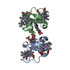
| Entry | Database: PDB / ID: 2abl | ||||||
|---|---|---|---|---|---|---|---|
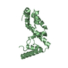
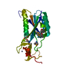
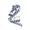
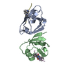
| Title | SH3-SH2 DOMAIN FRAGMENT OF HUMAN BCR-ABL TYROSINE KINASE | ||||||
 Components Components | ABL TYROSINE KINASE | ||||||
 Keywords Keywords | TRANSFERASE / TYROSINE KINASE / SH3 / SH2 / ONCOPROTEIN | ||||||
| Function / homology |  Function and homology information Function and homology informationnegative regulation of ubiquitin-protein transferase activity / protein localization to cytoplasmic microtubule plus-end / DNA conformation change / DN4 thymocyte differentiation / response to epinephrine / phospholipase C-inhibiting G protein-coupled receptor signaling pathway / podocyte apoptotic process / transitional one stage B cell differentiation / regulation of postsynaptic specialization assembly / positive regulation of phospholipase C/protein kinase C signal transduction ...negative regulation of ubiquitin-protein transferase activity / protein localization to cytoplasmic microtubule plus-end / DNA conformation change / DN4 thymocyte differentiation / response to epinephrine / phospholipase C-inhibiting G protein-coupled receptor signaling pathway / podocyte apoptotic process / transitional one stage B cell differentiation / regulation of postsynaptic specialization assembly / positive regulation of phospholipase C/protein kinase C signal transduction / regulation of modification of synaptic structure / nicotinate-nucleotide adenylyltransferase activity / delta-catenin binding / cerebellum morphogenesis / Role of ABL in ROBO-SLIT signaling / B cell proliferation involved in immune response / neuroepithelial cell differentiation / positive regulation of Wnt signaling pathway, planar cell polarity pathway / positive regulation of extracellular matrix organization / microspike assembly / B-1 B cell homeostasis / neuropilin signaling pathway / neuropilin binding / mitochondrial depolarization / regulation of cell motility / bubble DNA binding / positive regulation of establishment of T cell polarity / cellular response to dopamine / activated T cell proliferation / positive regulation of blood vessel branching / proline-rich region binding / negative regulation of mitotic cell cycle / mitogen-activated protein kinase binding / regulation of Cdc42 protein signal transduction / regulation of hematopoietic stem cell differentiation / syntaxin binding / positive regulation of dendrite development / regulation of axon extension / regulation of T cell differentiation / alpha-beta T cell differentiation / positive regulation of cell migration involved in sprouting angiogenesis / negative regulation of cell-cell adhesion / neuromuscular process controlling balance / Myogenesis / HDR through Single Strand Annealing (SSA) / positive regulation of osteoblast proliferation / platelet-derived growth factor receptor-beta signaling pathway / RUNX2 regulates osteoblast differentiation / Fc-gamma receptor signaling pathway involved in phagocytosis / vascular endothelial cell response to oscillatory fluid shear stress / Bergmann glial cell differentiation / regulation of endocytosis / regulation of microtubule polymerization / negative regulation of long-term synaptic potentiation / negative regulation of cellular senescence / myoblast proliferation / actin monomer binding / associative learning / positive regulation of focal adhesion assembly / negative regulation of BMP signaling pathway / positive regulation of vasoconstriction / ephrin receptor signaling pathway / cardiac muscle cell proliferation / BMP signaling pathway / RHO GTPases Activate WASPs and WAVEs / cellular response to transforming growth factor beta stimulus / negative regulation of endothelial cell apoptotic process / positive regulation of T cell migration / endothelial cell migration / negative regulation of double-strand break repair via homologous recombination / regulation of cell adhesion / positive regulation of interleukin-2 production / mismatch repair / ephrin receptor binding / spleen development / ERK1 and ERK2 cascade / four-way junction DNA binding / ruffle / positive regulation of stress fiber assembly / phosphotyrosine residue binding / signal transduction in response to DNA damage / canonical NF-kappaB signal transduction / actin filament polymerization / positive regulation of substrate adhesion-dependent cell spreading / substrate adhesion-dependent cell spreading / positive regulation of mitotic cell cycle / positive regulation of endothelial cell migration / establishment of localization in cell / SH2 domain binding / Turbulent (oscillatory, disturbed) flow shear stress activates signaling by PIEZO1 and integrins in endothelial cells / response to endoplasmic reticulum stress / thymus development / protein kinase C binding / protein modification process / positive regulation of release of sequestered calcium ion into cytosol / integrin-mediated signaling pathway / regulation of autophagy / protein serine/threonine kinase activator activity / B cell receptor signaling pathway / post-embryonic development Similarity search - Function | ||||||
| Biological species |  Homo sapiens (human) Homo sapiens (human) | ||||||
| Method |  X-RAY DIFFRACTION / MOLECULAR REPLACEMENT / Resolution: 2.5 Å X-RAY DIFFRACTION / MOLECULAR REPLACEMENT / Resolution: 2.5 Å | ||||||
 Authors Authors | Nam, H.-J. / Frederick, C.A. | ||||||
 Citation Citation | Journal: Structure / Year: 1996 Title: Intramolecular interactions of the regulatory domains of the Bcr-Abl kinase reveal a novel control mechanism. Authors: Nam, H.J. / Haser, W.G. / Roberts, T.M. / Frederick, C.A. | ||||||
| History |
|
- Structure visualization
| Structure viewer | Molecule: MolmilJmol/JSmol |
|---|
- Downloads & links
-Download
| PDBx/mmCIF format | 2abl.cif.gz | 45 KB | Display | PDBx/mmCIF format |
|---|---|---|---|---|
| PDB format | pdb2abl.ent.gz | 30.9 KB | Display | PDB format |
| PDBx/mmJSON format | 2abl.json.gz | Tree view | PDBx/mmJSON format | |
| Others |  Other downloads Other downloads |
-Validation report
| Arichive directory | https://data.pdbj.org/pub/pdb/validation_reports/ab/2ablftp://data.pdbj.org/pub/pdb/validation_reports/ab/2abl | HTTPS FTP |
|---|
-Related structure data
| Similar structure data |
|---|


-Links
 PDBj
PDBj


- Assembly
| Deposited unit | 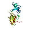
| ||||||||
|---|---|---|---|---|---|---|---|---|---|
| 1 |
| ||||||||
| Unit cell |
|
-Components
| #1: Protein | Mass: 18122.951 Da / Num. of mol.: 1 / Fragment: SH3-SH2 DOMAIN FRAGMENT / Mutation: INS(M76) Source method: isolated from a genetically manipulated source Source: (gene. exp.) Homo sapiens (human) / Cell line: BL21 / Gene: ABL SH3-SH2 / Plasmid: PRSET / Species (production host): Escherichia coli / Gene (production host): ABL SH3-SH2 / Production host:  |
|---|---|
| #2: Water | ChemComp-HOH /  Mass: 18.015 Da / Num. of mol.: 40 / Source method: isolated from a natural source / Formula: H2O Mass: 18.015 Da / Num. of mol.: 40 / Source method: isolated from a natural source / Formula: H2O |
-Experimental details
-Experiment
| Experiment | Method: X-RAY DIFFRACTION / Number of used crystals: 1 |
|---|
- Sample preparation
| Crystal | Density Matthews: 2.25 Å3/Da / Density % sol: 45.32 % | ||||||||||||||||||||||||||||||
|---|---|---|---|---|---|---|---|---|---|---|---|---|---|---|---|---|---|---|---|---|---|---|---|---|---|---|---|---|---|---|---|
| Crystal grow | pH: 7.5 / Details: pH 7.5 | ||||||||||||||||||||||||||||||
| Crystal grow | *PLUS Method: vapor diffusion, hanging dropDetails: drop solution was mixed with an equal volume of mother liquor | ||||||||||||||||||||||||||||||
| Components of the solutions | *PLUS
|
-Data collection
| Diffraction | Mean temperature: 293 K |
|---|---|
| Diffraction source | Source: ROTATING ANODE / Type: RIGAKU RUH3R / Wavelength: 1.5418 |
| Detector | Type: MARRESEARCH / Detector: IMAGE PLATE / Date: Nov 1, 1993 / Details: MIRRORS |
| Radiation | Monochromator: NI FILTER / Monochromatic (M) / Laue (L): M / Scattering type: x-ray |
| Radiation wavelength | Wavelength: 1.5418 Å / Relative weight: 1 |
| Reflection | Resolution: 2.5→12 Å / Num. obs: 5585 / % possible obs: 99.3 % / Redundancy: 3.5 % / Rsym value: 0.048 |
| Reflection shell | Resolution: 2.5→2.59 Å / Redundancy: 3.3 % / Rsym value: 0.14 / % possible all: 98 |
| Reflection | *PLUS Num. measured all: 19460 / Rmerge(I) obs: 0.048 |
| Reflection shell | *PLUS % possible obs: 98 % / Rmerge(I) obs: 0.14 |
- Processing
| Software |
| ||||||||||||||||||||||||||||||||||||||||||||||||||||||||||||
|---|---|---|---|---|---|---|---|---|---|---|---|---|---|---|---|---|---|---|---|---|---|---|---|---|---|---|---|---|---|---|---|---|---|---|---|---|---|---|---|---|---|---|---|---|---|---|---|---|---|---|---|---|---|---|---|---|---|---|---|---|---|
| Refinement | Method to determine structure: MOLECULAR REPLACEMENT Starting model: ABL SH3, SRC SH2 Resolution: 2.5→10 Å / Cross valid method: FREE-R / σ(F): 2
| ||||||||||||||||||||||||||||||||||||||||||||||||||||||||||||
| Refinement step | Cycle: LAST / Resolution: 2.5→10 Å
| ||||||||||||||||||||||||||||||||||||||||||||||||||||||||||||
| Refine LS restraints |
| ||||||||||||||||||||||||||||||||||||||||||||||||||||||||||||
| Software | *PLUS Name: X-PLOR / Version: 3 / Classification: refinement | ||||||||||||||||||||||||||||||||||||||||||||||||||||||||||||
| Refinement | *PLUS | ||||||||||||||||||||||||||||||||||||||||||||||||||||||||||||
| Solvent computation | *PLUS | ||||||||||||||||||||||||||||||||||||||||||||||||||||||||||||
| Displacement parameters | *PLUS | ||||||||||||||||||||||||||||||||||||||||||||||||||||||||||||
| Refine LS restraints | *PLUS
|
