 Movie
Movie Controller

[English] 日本語
 Yorodumi
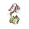
- PDB-1cei: STRUCTURE DETERMINATION OF THE COLICIN E7 IMMUNITY PROTEIN (IMME7... -
+ Open data

- Basic information
| Entry | Database: PDB / ID: 1cei | ||||||
|---|---|---|---|---|---|---|---|
| Title | STRUCTURE DETERMINATION OF THE COLICIN E7 IMMUNITY PROTEIN (IMME7) THAT BINDS SPECIFICALLY TO THE DNASE-TYPE COLICIN E7 AND INHIBITS ITS BACTERIOCIDAL ACTIVITY | ||||||
 Components Components | COLICIN E7 IMMUNITY PROTEIN | ||||||
 Keywords Keywords | ANTIBACTERIAL PROTEIN / IMMUNITY PROTEIN | ||||||
| Function / homology |  Function and homology information Function and homology information | ||||||
| Biological species |  | ||||||
| Method |  X-RAY DIFFRACTION / MIR / Resolution: 1.8 Å X-RAY DIFFRACTION / MIR / Resolution: 1.8 Å | ||||||
 Authors Authors | Chak, K.-F. / Safo, M.K. / Ku, W.-Y. / Hsieh, S.-Y. / Yuan, H.S. | ||||||
 Citation Citation | Journal: Proc.Natl.Acad.Sci.USA / Year: 1996 Title: The crystal structure of the immunity protein of colicin E7 suggests a possible colicin-interacting surface. Authors: Chak, K.F. / Safo, M.K. / Ku, W.Y. / Hsieh, S.Y. / Yuan, H.S. | ||||||
| History |
|
- Structure visualization
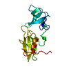
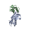
| Structure viewer | Molecule: MolmilJmol/JSmol |
|---|
- Downloads & links
-Download
| PDBx/mmCIF format | 1cei.cif.gz | 28.2 KB | Display | PDBx/mmCIF format |
|---|---|---|---|---|
| PDB format | pdb1cei.ent.gz | 18.4 KB | Display | PDB format |
| PDBx/mmJSON format | 1cei.json.gz | Tree view | PDBx/mmJSON format | |
| Others |  Other downloads Other downloads |
-Validation report
| Arichive directory | https://data.pdbj.org/pub/pdb/validation_reports/ce/1ceiftp://data.pdbj.org/pub/pdb/validation_reports/ce/1cei | HTTPS FTP |
|---|
-Related structure data
| Similar structure data |
|---|


-Links
 PDBj
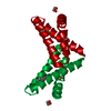
PDBj- Assembly
| Deposited unit | 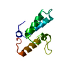
| ||||||||
|---|---|---|---|---|---|---|---|---|---|
| 1 | 
| ||||||||
| Unit cell |
| ||||||||
| Details | THERE IS ONE MONOMER IN THE ASYMMETRIC UNIT. THE FIRST TWO N-TERMINAL RESIDUES ARE DISORDERED AND ARE NOT OBSERVED IN THE ELECTRON DENSITY MAP. |
-Components
| #1: Protein | Mass: 10651.846 Da / Num. of mol.: 1 / Source method: isolated from a natural source / Source: (natural) |
|---|---|
| #2: Water | ChemComp-HOH /  Mass: 18.015 Da / Num. of mol.: 60 / Source method: isolated from a natural source / Formula: H2O Mass: 18.015 Da / Num. of mol.: 60 / Source method: isolated from a natural source / Formula: H2O |
-Experimental details
-Experiment
| Experiment | Method: X-RAY DIFFRACTION / Number of used crystals: 1 |
|---|
- Sample preparation
| Crystal | Density Matthews: 2.02 Å3/Da / Density % sol: 39.05 % | ||||||||||||||||||||
|---|---|---|---|---|---|---|---|---|---|---|---|---|---|---|---|---|---|---|---|---|---|
| Crystal grow | pH: 8 / Details: pH 8. | ||||||||||||||||||||
| Crystal | *PLUS Density % sol: 43 % | ||||||||||||||||||||
| Crystal grow | *PLUS Method: vapor diffusion, hanging drop | ||||||||||||||||||||
| Components of the solutions | *PLUS
|
-Data collection
| Diffraction | Mean temperature: 298 K |
|---|---|
| Diffraction source | Source: ROTATING ANODE / Type: RIGAKU RUH3R / Wavelength: 1.5418 |
| Detector | Type: RIGAKU RAXIS II / Detector: IMAGE PLATE / Date: Nov 21, 1994 |
| Radiation | Monochromatic (M) / Laue (L): M / Scattering type: x-ray |
| Radiation wavelength | Wavelength: 1.5418 Å / Relative weight: 1 |
| Reflection | Resolution: 1.8→50 Å / Num. obs: 7917 / % possible obs: 95.2 % / Observed criterion σ(I): 2 / Redundancy: 3.97 % / Rmerge(I) obs: 0.054 |
| Reflection shell | Resolution: 1.8→1.9 Å / Redundancy: 3.2 % / Rmerge(I) obs: 0.21 / Mean I/σ(I) obs: 2.9 / % possible all: 78.1 |
| Reflection | *PLUS Num. measured all: 31416 |
| Reflection shell | *PLUS % possible obs: 78.1 % |
- Processing
| Software |
| ||||||||||||||||||||||||||||||||||||||||||||||||||||||||||||
|---|---|---|---|---|---|---|---|---|---|---|---|---|---|---|---|---|---|---|---|---|---|---|---|---|---|---|---|---|---|---|---|---|---|---|---|---|---|---|---|---|---|---|---|---|---|---|---|---|---|---|---|---|---|---|---|---|---|---|---|---|---|
| Refinement | Method to determine structure: MIR / Resolution: 1.8→6 Å / σ(F): 2
| ||||||||||||||||||||||||||||||||||||||||||||||||||||||||||||
| Displacement parameters | Biso mean: 28.5 Å2 | ||||||||||||||||||||||||||||||||||||||||||||||||||||||||||||
| Refine analyze | Luzzati coordinate error obs: 0.22 Å | ||||||||||||||||||||||||||||||||||||||||||||||||||||||||||||
| Refinement step | Cycle: LAST / Resolution: 1.8→6 Å
| ||||||||||||||||||||||||||||||||||||||||||||||||||||||||||||
| Refine LS restraints |
| ||||||||||||||||||||||||||||||||||||||||||||||||||||||||||||
| Software | *PLUS Name: X-PLOR / Classification: refinement | ||||||||||||||||||||||||||||||||||||||||||||||||||||||||||||
| Refinement | *PLUS | ||||||||||||||||||||||||||||||||||||||||||||||||||||||||||||
| Solvent computation | *PLUS | ||||||||||||||||||||||||||||||||||||||||||||||||||||||||||||
| Displacement parameters | *PLUS | ||||||||||||||||||||||||||||||||||||||||||||||||||||||||||||
| Refine LS restraints | *PLUS
|
