 Movie
Movie Controller

[English] 日本語
 Yorodumi
- PDB-1a02: STRUCTURE OF THE DNA BINDING DOMAINS OF NFAT, FOS AND JUN BOUND TO DNA -
+ Open data

- Basic information
| Entry | Database: PDB / ID: 1a02 | ||||||
|---|---|---|---|---|---|---|---|
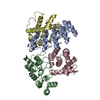
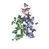
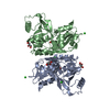
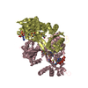
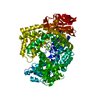
| Title | STRUCTURE OF THE DNA BINDING DOMAINS OF NFAT, FOS AND JUN BOUND TO DNA | ||||||
 Components Components |
| ||||||
 Keywords Keywords | TRANSCRIPTION/DNA / TRANSCRIPTION FACTOR / NFAT / NF-AT / AP-1 / FOS-JUN / QUATERNARY PROTEIN-DNA COMPLEX / TRANSCRIPTION SYNERGY / COMBINATORIAL GENE REGULATION / TRANSCRIPTION-DNA COMPLEX | ||||||
| Function / homology |  Function and homology information Function and homology informationcellular response to prolactin / medium-term memory / lncRNA transcription / response to forskolin / leading edge cell differentiation / cellular response to anisomycin / cAMP response element binding / transcription factor AP-1 complex / mononuclear cell differentiation / negative regulation of vascular associated smooth muscle cell differentiation ...cellular response to prolactin / medium-term memory / lncRNA transcription / response to forskolin / leading edge cell differentiation / cellular response to anisomycin / cAMP response element binding / transcription factor AP-1 complex / mononuclear cell differentiation / negative regulation of vascular associated smooth muscle cell differentiation / integrated stress response signaling / cellular response to zinc ion starvation / negative regulation of DNA binding / release from viral latency / skeletal muscle cell proliferation / myotube cell development / RUNX1 and FOXP3 control the development of regulatory T lymphocytes (Tregs) / calcineurin-NFAT signaling cascade / WNT5:FZD7-mediated leishmania damping / neural retina development / cellular response to phorbol 13-acetate 12-myristate / SMAD protein signal transduction / NGF-stimulated transcription / conditioned taste aversion / positive regulation of osteoclast differentiation / host-mediated activation of viral transcription / cartilage development / response to steroid hormone / axon regeneration / positive regulation of DNA-templated transcription initiation / positive regulation of myoblast fusion / cellular response to parathyroid hormone stimulus / Deregulated CDK5 triggers multiple neurodegenerative pathways in Alzheimer's disease models / CLEC7A (Dectin-1) induces NFAT activation / response to corticosterone / Activation of the AP-1 family of transcription factors / nuclear chromosome / eyelid development in camera-type eye / response to immobilization stress / response to gravity / outflow tract morphogenesis / R-SMAD binding / positive regulation of epithelial cell migration / myoblast proliferation / ubiquitin-like protein ligase binding / host-mediated suppression of viral transcription / Calcineurin activates NFAT / response to light stimulus / skeletal muscle cell differentiation / monocyte differentiation / Estrogen-dependent nuclear events downstream of ESR-membrane signaling / general transcription initiation factor binding / response to cAMP / RNA polymerase II core promoter sequence-specific DNA binding / phosphatase binding / positive regulation of vascular associated smooth muscle cell proliferation / JNK cascade / NPAS4 regulates expression of target genes / positive regulation of B cell proliferation / response to muscle stretch / transcription repressor complex / response to progesterone / positive regulation of endothelial cell proliferation / 14-3-3 protein binding / FCERI mediated Ca+2 mobilization / transforming growth factor beta receptor signaling pathway / osteoclast differentiation / cellular response to calcium ion / cellular response to epidermal growth factor stimulus / response to endoplasmic reticulum stress / response to activity / Regulation of PTEN gene transcription / transcription coregulator binding / GTPase activator activity / B cell receptor signaling pathway / FCERI mediated MAPK activation / TP53 Regulates Transcription of DNA Repair Genes / cellular response to reactive oxygen species / liver development / promoter-specific chromatin binding / female pregnancy / euchromatin / microglial cell activation / protein-DNA complex / cerebral cortex development / response to insulin / positive regulation of miRNA transcription / DNA-binding transcription repressor activity, RNA polymerase II-specific / MAPK6/MAPK4 signaling / Pre-NOTCH Transcription and Translation / cellular response to tumor necrosis factor / RNA polymerase II transcription regulator complex / positive regulation of fibroblast proliferation / Activation of anterior HOX genes in hindbrain development during early embryogenesis / response to toxic substance / neuron differentiation / nuclear matrix / sequence-specific double-stranded DNA binding / Signaling by ALK fusions and activated point mutants / regulation of cell population proliferation Similarity search - Function | ||||||
| Biological species |  Homo sapiens (human) Homo sapiens (human) | ||||||
| Method |  X-RAY DIFFRACTION / SYNCHROTRON / MIR/MAD / Resolution: 2.7 Å X-RAY DIFFRACTION / SYNCHROTRON / MIR/MAD / Resolution: 2.7 Å | ||||||
 Authors Authors | Chen, L. / Glover, J.N.M. / Hogan, P.G. / Rao, A. / Harrison, S.C. | ||||||
 Citation Citation | Journal: Nature / Year: 1998 Title: Structure of the DNA-binding domains from NFAT, Fos and Jun bound specifically to DNA. Authors: Chen, L. / Glover, J.N. / Hogan, P.G. / Rao, A. / Harrison, S.C. | ||||||
| History |
|
- Structure visualization
| Structure viewer | Molecule: MolmilJmol/JSmol |
|---|
- Downloads & links
-Download
| PDBx/mmCIF format | 1a02.cif.gz | 114.8 KB | Display | PDBx/mmCIF format |
|---|---|---|---|---|
| PDB format | pdb1a02.ent.gz | 84.7 KB | Display | PDB format |
| PDBx/mmJSON format | 1a02.json.gz | Tree view | PDBx/mmJSON format | |
| Others |  Other downloads Other downloads |
-Validation report
| Arichive directory | https://data.pdbj.org/pub/pdb/validation_reports/a0/1a02ftp://data.pdbj.org/pub/pdb/validation_reports/a0/1a02 | HTTPS FTP |
|---|
-Related structure data
| Similar structure data |
|---|


-Links
 PDBj
PDBj


- Assembly
| Deposited unit | 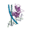
| ||||||||||
|---|---|---|---|---|---|---|---|---|---|---|---|
| 1 |
| ||||||||||
| Unit cell |
|
-Components
-DNA chain , 2 types, 2 molecules AB
| #1: DNA chain | Mass: 6178.024 Da / Num. of mol.: 1 / Source method: obtained synthetically |
|---|---|
| #2: DNA chain | Mass: 6084.993 Da / Num. of mol.: 1 / Source method: obtained synthetically |
-AP-1 FRAGMENT ... , 2 types, 2 molecules FJ
| #4: Protein | Mass: 6727.733 Da / Num. of mol.: 1 / Fragment: FOS / Mutation: C154S Source method: isolated from a genetically manipulated source Source: (gene. exp.) Homo sapiens (human) / Production host:  |
|---|---|
| #5: Protein | Mass: 6583.826 Da / Num. of mol.: 1 / Fragment: JUN / Mutation: C279S Source method: isolated from a genetically manipulated source Source: (gene. exp.) Homo sapiens (human) / Production host: |
-Protein / Non-polymers , 2 types, 89 molecules N
| #3: Protein | Mass: 34270.898 Da / Num. of mol.: 1 Source method: isolated from a genetically manipulated source Source: (gene. exp.) Homo sapiens (human) / Cell: T-LYMPHOCYTE / Gene: NFAT1 / Plasmid: PLM1 / Species (production host): Escherichia coli / Production host: |
|---|---|
| #6: Water | ChemComp-HOH / Mass: 18.015 Da / Num. of mol.: 88 / Source method: isolated from a natural source / Formula: H2O |
-Experimental details
-Experiment
| Experiment | Method: X-RAY DIFFRACTION / Number of used crystals: 1 |
|---|
- Sample preparation
| Crystal | Density Matthews: 3.54 Å3/Da / Density % sol: 68 % | ||||||||||||||||||||||||||||||||||||||||||||||||||||||||||||||||||
|---|---|---|---|---|---|---|---|---|---|---|---|---|---|---|---|---|---|---|---|---|---|---|---|---|---|---|---|---|---|---|---|---|---|---|---|---|---|---|---|---|---|---|---|---|---|---|---|---|---|---|---|---|---|---|---|---|---|---|---|---|---|---|---|---|---|---|---|
| Crystal grow | Method: vapor diffusion, hanging drop / pH: 7.5 Details: THE COMPLEX WAS CRYSTALLIZED IN 300-400 MM AMMONIUM ACETATE SALT, PH 7.5 (10 MM)., VAPOR DIFFUSION, HANGING DROP | ||||||||||||||||||||||||||||||||||||||||||||||||||||||||||||||||||
| Components of the solutions |
| ||||||||||||||||||||||||||||||||||||||||||||||||||||||||||||||||||
| Crystal | *PLUS Density % sol: 68 % | ||||||||||||||||||||||||||||||||||||||||||||||||||||||||||||||||||
| Crystal grow | *PLUS | ||||||||||||||||||||||||||||||||||||||||||||||||||||||||||||||||||
| Components of the solutions | *PLUS
|
-Data collection
| Diffraction | Mean temperature: 100 K |
|---|---|
| Diffraction source | Source: SYNCHROTRON / Site: NSLS  / Beamline: X25 / Beamline: X25 |
| Detector | Type: MARRESEARCH / Detector: IMAGE PLATE / Date: Sep 16, 1996 |
| Radiation | Protocol: SINGLE WAVELENGTH / Monochromatic (M) / Laue (L): M / Scattering type: x-ray |
| Radiation wavelength | Relative weight: 1 |
| Reflection | Resolution: 2.7→20 Å / Num. obs: 22079 / % possible obs: 98.3 % / Observed criterion σ(I): 0 / Redundancy: 3.1 % / Biso Wilson estimate: 61 Å2 / Rsym value: 0.08 |
| Reflection shell | Resolution: 2.7→2.8 Å / Redundancy: 2.7 % / Rsym value: 0.43 / % possible all: 93.3 |
| Reflection | *PLUS Highest resolution: 2.7 Å / Lowest resolution: 20 Å / % possible obs: 98.3 % / Redundancy: 3.1 % / Rmerge(I) obs: 0.08 / Biso Wilson estimate: 61 Å2 |
| Reflection shell | *PLUS Highest resolution: 2.7 Å / Lowest resolution: 2.8 Å / % possible obs: 93.3 % / Rmerge(I) obs: 0.43 / Mean I/σ(I) obs: 2.6 |
- Processing
| Software |
| ||||||||||||||||||||||||||||||||||||||||||||||||||||||||||||||||||||||||||||||||
|---|---|---|---|---|---|---|---|---|---|---|---|---|---|---|---|---|---|---|---|---|---|---|---|---|---|---|---|---|---|---|---|---|---|---|---|---|---|---|---|---|---|---|---|---|---|---|---|---|---|---|---|---|---|---|---|---|---|---|---|---|---|---|---|---|---|---|---|---|---|---|---|---|---|---|---|---|---|---|---|---|---|
| Refinement | Method to determine structure: MIR/MAD / Resolution: 2.7→10 Å / Rfactor Rfree error: 0.01 / Data cutoff high absF: 10000000 / Data cutoff low absF: 0.1 / Isotropic thermal model: RESTRAINED / Cross valid method: THROUGHOUT / σ(F): 2 Details: RESIDUES N 478 - N 485 AND N 628 - N 634 ARE DISORDERED
| ||||||||||||||||||||||||||||||||||||||||||||||||||||||||||||||||||||||||||||||||
| Displacement parameters | Biso mean: 51 Å2 | ||||||||||||||||||||||||||||||||||||||||||||||||||||||||||||||||||||||||||||||||
| Refine analyze | Luzzati d res low obs: 10 Å | ||||||||||||||||||||||||||||||||||||||||||||||||||||||||||||||||||||||||||||||||
| Refinement step | Cycle: LAST / Resolution: 2.7→10 Å
| ||||||||||||||||||||||||||||||||||||||||||||||||||||||||||||||||||||||||||||||||
| Refine LS restraints |
| ||||||||||||||||||||||||||||||||||||||||||||||||||||||||||||||||||||||||||||||||
| LS refinement shell | Resolution: 2.7→2.82 Å / Rfactor Rfree error: 0.02 / Total num. of bins used: 8
| ||||||||||||||||||||||||||||||||||||||||||||||||||||||||||||||||||||||||||||||||
| Xplor file |
| ||||||||||||||||||||||||||||||||||||||||||||||||||||||||||||||||||||||||||||||||
| Software | *PLUS Name: X-PLOR / Version: 3.1 / Classification: refinement | ||||||||||||||||||||||||||||||||||||||||||||||||||||||||||||||||||||||||||||||||
| Refinement | *PLUS Highest resolution: 2.7 Å / Lowest resolution: 10 Å / σ(F): 2 / % reflection Rfree: 7.5 % | ||||||||||||||||||||||||||||||||||||||||||||||||||||||||||||||||||||||||||||||||
| Solvent computation | *PLUS | ||||||||||||||||||||||||||||||||||||||||||||||||||||||||||||||||||||||||||||||||
| Displacement parameters | *PLUS | ||||||||||||||||||||||||||||||||||||||||||||||||||||||||||||||||||||||||||||||||
| Refine LS restraints | *PLUS
| ||||||||||||||||||||||||||||||||||||||||||||||||||||||||||||||||||||||||||||||||
| LS refinement shell | *PLUS Rfactor obs: 0.369 |
