 Movie
Movie Controller

[English] 日本語
 Yorodumi
- PDB-2rc7: Crystal structure of the NR3A ligand binding core complex with gl... -
+ Open data

- Basic information
| Entry | Database: PDB / ID: 2rc7 | ||||||
|---|---|---|---|---|---|---|---|
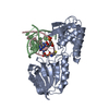
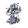
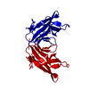
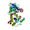
| Title | Crystal structure of the NR3A ligand binding core complex with glycine at 1.58 Angstrom resolution | ||||||
 Components Components | Glutamate [NMDA] receptor subunit 3A | ||||||
 Keywords Keywords | MEMBRANE PROTEIN / Cell junction / Glycoprotein / Ion transport / Ionic channel / Magnesium / Postsynaptic cell membrane / Receptor / Synapse / Transmembrane / Transport | ||||||
| Function / homology |  Function and homology information Function and homology informationserine binding / negative regulation of dendritic spine development / Assembly and cell surface presentation of NMDA receptors / transmitter-gated monoatomic ion channel activity / glutamate receptor activity / neurotransmitter receptor complex / NMDA glutamate receptor activity / NMDA selective glutamate receptor complex / glycine binding / dendrite development ...serine binding / negative regulation of dendritic spine development / Assembly and cell surface presentation of NMDA receptors / transmitter-gated monoatomic ion channel activity / glutamate receptor activity / neurotransmitter receptor complex / NMDA glutamate receptor activity / NMDA selective glutamate receptor complex / glycine binding / dendrite development / monoatomic ion channel complex / monoatomic cation transmembrane transport / neuron development / prepulse inhibition / glutamate-gated receptor activity / ionotropic glutamate receptor signaling pathway / protein phosphatase 2A binding / presynaptic modulation of chemical synaptic transmission / synaptic transmission, glutamatergic / transmitter-gated monoatomic ion channel activity involved in regulation of postsynaptic membrane potential / regulation of synaptic plasticity / postsynaptic density membrane / modulation of chemical synaptic transmission / calcium channel activity / calcium ion transport / rhythmic process / presynapse / response to ethanol / postsynaptic membrane / neuron projection / neuronal cell body / synapse / endoplasmic reticulum membrane / glutamatergic synapse / membrane / identical protein binding / plasma membrane / cytoplasm Similarity search - Function | ||||||
| Biological species |  | ||||||
| Method |  X-RAY DIFFRACTION / SYNCHROTRON / MOLECULAR REPLACEMENT / Resolution: 1.58 Å X-RAY DIFFRACTION / SYNCHROTRON / MOLECULAR REPLACEMENT / Resolution: 1.58 Å | ||||||
 Authors Authors | Yao, Y. / Mayer, M.L. | ||||||
 Citation Citation | Journal: Embo J. / Year: 2008 Title: Molecular mechanism of ligand recognition by NR3 subtype glutamate receptors. Authors: Yao, Y. / Harrison, C.B. / Freddolino, P.L. / Schulten, K. / Mayer, M.L. #1: Journal: J.NEUROSCI. / Year: 2006 Title: Characterization of a soluble ligand binding domain of the NMDA receptor regulatory subunit NR3A Authors: Yao, Y. / Mayer, M.L. | ||||||
| History |
| ||||||
| Remark 999 | RESIDUES 153 AND 154, GLY AND THR, ARE INSERTED AS A LINK REPLACING RESIDUES 661-775 OF THE PROTEIN ...RESIDUES 153 AND 154, GLY AND THR, ARE INSERTED AS A LINK REPLACING RESIDUES 661-775 OF THE PROTEIN FROM THE UNP entry Q9R1M7 |
- Structure visualization
| Structure viewer | Molecule: MolmilJmol/JSmol |
|---|
- Downloads & links
-Download
| PDBx/mmCIF format | 2rc7.cif.gz | 148.5 KB | Display | PDBx/mmCIF format |
|---|---|---|---|---|
| PDB format | pdb2rc7.ent.gz | 116.7 KB | Display | PDB format |
| PDBx/mmJSON format | 2rc7.json.gz | Tree view | PDBx/mmJSON format | |
| Others |  Other downloads Other downloads |
-Validation report
| Arichive directory | https://data.pdbj.org/pub/pdb/validation_reports/rc/2rc7ftp://data.pdbj.org/pub/pdb/validation_reports/rc/2rc7 | HTTPS FTP |
|---|
-Related structure data
| Related structure data |  2rc8C  2rc9C  2rcaC  2rcbC  1pb7S C: citing same article ( S: Starting model for refinement |
|---|---|
| Similar structure data |


-Links
 PDBj
PDBj


- Assembly
| Deposited unit | 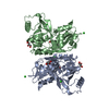
| ||||||||
|---|---|---|---|---|---|---|---|---|---|
| 1 | 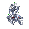
| ||||||||
| 2 | 
| ||||||||
| Unit cell |
| ||||||||
| Details | The biological unit is believed to be a dimer of dimers. Molecular packing in the present structure is not biologically relevant. |
-Components
-Protein , 1 types, 2 molecules AB
| #1: Protein | Mass: 32936.301 Da / Num. of mol.: 2 / Fragment: unp residues 511-660, 776-915 Source method: isolated from a genetically manipulated source Source: (gene. exp.) Description: Peptides corresponding to N511-R660 and E776-K915 were coupled by a GT dipeptid e synthetic linker Gene: Grin3a / Plasmid: pET22b(+) modified / Production host:  |
|---|
-Non-polymers , 5 types, 645 molecules 


| #2: Chemical | ChemComp-BR /  Mass: 79.904 Da / Num. of mol.: 1 / Source method: obtained synthetically / Formula: Br Mass: 79.904 Da / Num. of mol.: 1 / Source method: obtained synthetically / Formula: Br | ||||||
|---|---|---|---|---|---|---|---|
| #3: Chemical | ChemComp-CL /  Mass: 35.453 Da / Num. of mol.: 7 / Source method: obtained synthetically / Formula: Cl Mass: 35.453 Da / Num. of mol.: 7 / Source method: obtained synthetically / Formula: Cl#4: Chemical | ChemComp-GOL /  Mass: 92.094 Da / Num. of mol.: 4 / Source method: obtained synthetically / Formula: C3H8O3 Mass: 92.094 Da / Num. of mol.: 4 / Source method: obtained synthetically / Formula: C3H8O3#5: Chemical |  Type: peptide linking / Mass: 75.067 Da / Num. of mol.: 2 / Source method: obtained synthetically / Formula: C2H5NO2 Type: peptide linking / Mass: 75.067 Da / Num. of mol.: 2 / Source method: obtained synthetically / Formula: C2H5NO2#6: Water | ChemComp-HOH / | Mass: 18.015 Da / Num. of mol.: 631 / Source method: isolated from a natural source / Formula: H2O |
-Details
| Has protein modification | Y |
|---|
-Experimental details
-Experiment
| Experiment | Method: X-RAY DIFFRACTION / Number of used crystals: 1 |
|---|
- Sample preparation
| Crystal | Density Matthews: 2.21 Å3/Da / Density % sol: 44.32 % |
|---|---|
| Crystal grow | Temperature: 293 K / Method: vapor diffusion, hanging drop / pH: 5.2 Details: 0.1 M NaBr, 0.1 mM NaAcetate, 4% PEG 4000, pH 5.2, VAPOR DIFFUSION, HANGING DROP, temperature 293K |
-Data collection
| Diffraction | Mean temperature: 100 K |
|---|---|
| Diffraction source | Source: SYNCHROTRON / Site: APS  / Beamline: 22-ID / Wavelength: 0.97931 Å / Beamline: 22-ID / Wavelength: 0.97931 Å |
| Detector | Type: MARMOSAIC 300 mm CCD / Detector: CCD / Date: Mar 25, 2006 |
| Radiation | Monochromator: Si 220 / Protocol: SINGLE WAVELENGTH / Monochromatic (M) / Laue (L): M / Scattering type: x-ray |
| Radiation wavelength | Wavelength: 0.97931 Å / Relative weight: 1 |
| Reflection | Resolution: 1.58→30 Å / Num. all: 74941 / Num. obs: 74941 / % possible obs: 95.9 % / Observed criterion σ(F): 1 / Observed criterion σ(I): 1 / Redundancy: 3.8 % / Rmerge(I) obs: 0.054 / Net I/σ(I): 11.7 |
| Reflection shell | Resolution: 1.58→1.64 Å / Redundancy: 3.3 % / Rmerge(I) obs: 0.209 / Mean I/σ(I) obs: 5.6 / % possible all: 75.3 |
- Processing
| Software |
| |||||||||||||||||||||||||||||||||||||||||||||||||||||||||||||||||||||||||||||||||||||||||||||||||||||||||||||||||||||||||||||||||||||||||||||||||||||||||||||||||||||||||||||||
|---|---|---|---|---|---|---|---|---|---|---|---|---|---|---|---|---|---|---|---|---|---|---|---|---|---|---|---|---|---|---|---|---|---|---|---|---|---|---|---|---|---|---|---|---|---|---|---|---|---|---|---|---|---|---|---|---|---|---|---|---|---|---|---|---|---|---|---|---|---|---|---|---|---|---|---|---|---|---|---|---|---|---|---|---|---|---|---|---|---|---|---|---|---|---|---|---|---|---|---|---|---|---|---|---|---|---|---|---|---|---|---|---|---|---|---|---|---|---|---|---|---|---|---|---|---|---|---|---|---|---|---|---|---|---|---|---|---|---|---|---|---|---|---|---|---|---|---|---|---|---|---|---|---|---|---|---|---|---|---|---|---|---|---|---|---|---|---|---|---|---|---|---|---|---|---|---|
| Refinement | Method to determine structure: MOLECULAR REPLACEMENT Starting model: Pdb entry 1PB7 Resolution: 1.58→29.89 Å / Cor.coef. Fo:Fc: 0.968 / Cor.coef. Fo:Fc free: 0.957 / SU B: 2.52 / SU ML: 0.048 / TLS residual ADP flag: LIKELY RESIDUAL / Cross valid method: THROUGHOUT / σ(F): 0 / ESU R: 0.081 / ESU R Free: 0.081 / Stereochemistry target values: MAXIMUM LIKELIHOOD / Details: HYDROGENS HAVE BEEN ADDED IN THE RIDING POSITIONS
| |||||||||||||||||||||||||||||||||||||||||||||||||||||||||||||||||||||||||||||||||||||||||||||||||||||||||||||||||||||||||||||||||||||||||||||||||||||||||||||||||||||||||||||||
| Solvent computation | Ion probe radii: 0.8 Å / Shrinkage radii: 0.8 Å / VDW probe radii: 1.4 Å / Solvent model: BABINET MODEL WITH MASK | |||||||||||||||||||||||||||||||||||||||||||||||||||||||||||||||||||||||||||||||||||||||||||||||||||||||||||||||||||||||||||||||||||||||||||||||||||||||||||||||||||||||||||||||
| Displacement parameters | Biso mean: 13.485 Å2
| |||||||||||||||||||||||||||||||||||||||||||||||||||||||||||||||||||||||||||||||||||||||||||||||||||||||||||||||||||||||||||||||||||||||||||||||||||||||||||||||||||||||||||||||
| Refinement step | Cycle: LAST / Resolution: 1.58→29.89 Å
| |||||||||||||||||||||||||||||||||||||||||||||||||||||||||||||||||||||||||||||||||||||||||||||||||||||||||||||||||||||||||||||||||||||||||||||||||||||||||||||||||||||||||||||||
| Refine LS restraints |
| |||||||||||||||||||||||||||||||||||||||||||||||||||||||||||||||||||||||||||||||||||||||||||||||||||||||||||||||||||||||||||||||||||||||||||||||||||||||||||||||||||||||||||||||
| LS refinement shell | Resolution: 1.58→1.621 Å / Total num. of bins used: 20
| |||||||||||||||||||||||||||||||||||||||||||||||||||||||||||||||||||||||||||||||||||||||||||||||||||||||||||||||||||||||||||||||||||||||||||||||||||||||||||||||||||||||||||||||
| Refinement TLS params. | Method: refined / Refine-ID: X-RAY DIFFRACTION
| |||||||||||||||||||||||||||||||||||||||||||||||||||||||||||||||||||||||||||||||||||||||||||||||||||||||||||||||||||||||||||||||||||||||||||||||||||||||||||||||||||||||||||||||
| Refinement TLS group |
|
