[English] 日本語
Yorodumi
- PDB-4kcd: Crystal Structure of the NMDA Receptor GluN3A Ligand Binding Doma... -

+
Open data


ID or keywords:

Loading...

-
Basic information

Entry
Database: PDB / ID: 4kcd
TitleCrystal Structure of the NMDA Receptor GluN3A Ligand Binding Domain Apo State
ComponentsGlutamate receptor ionotropic, NMDA 3A
KeywordsMEMBRANE PROTEIN
Function / homology
Function and homology information


negative regulation of dendritic spine development / Assembly and cell surface presentation of NMDA receptors / glutamate receptor activity / NMDA glutamate receptor activity / NMDA selective glutamate receptor complex / glycine binding / dendrite development / neuron development / prepulse inhibition / glutamate-gated receptor activity ...negative regulation of dendritic spine development / Assembly and cell surface presentation of NMDA receptors / glutamate receptor activity / NMDA glutamate receptor activity / NMDA selective glutamate receptor complex / glycine binding / dendrite development / neuron development / prepulse inhibition / glutamate-gated receptor activity / presynaptic modulation of chemical synaptic transmission / ionotropic glutamate receptor signaling pathway / protein phosphatase 2A binding / synaptic transmission, glutamatergic / transmitter-gated monoatomic ion channel activity involved in regulation of postsynaptic membrane potential / postsynaptic density membrane / calcium channel activity / modulation of chemical synaptic transmission / calcium ion transport / rhythmic process / presynapse / postsynaptic membrane / response to ethanol / neuron projection / neuronal cell body / glutamatergic synapse / synapse / endoplasmic reticulum membrane / identical protein binding / membrane / plasma membrane / cytoplasm
Similarity search - Function
Bacterial extracellular solute-binding proteins, family 3 / Solute-binding protein family 3/N-terminal domain of MltF / Ionotropic glutamate receptor, metazoa / Ligated ion channel L-glutamate- and glycine-binding site / : / Ligand-gated ion channel / Ionotropic glutamate receptor, L-glutamate and glycine-binding domain / Ligated ion channel L-glutamate- and glycine-binding site / Ionotropic glutamate receptor / Eukaryotic homologues of bacterial periplasmic substrate binding proteins. ...Bacterial extracellular solute-binding proteins, family 3 / Solute-binding protein family 3/N-terminal domain of MltF / Ionotropic glutamate receptor, metazoa / Ligated ion channel L-glutamate- and glycine-binding site / : / Ligand-gated ion channel / Ionotropic glutamate receptor, L-glutamate and glycine-binding domain / Ligated ion channel L-glutamate- and glycine-binding site / Ionotropic glutamate receptor / Eukaryotic homologues of bacterial periplasmic substrate binding proteins. / Receptor, ligand binding region / Receptor family ligand binding region / Periplasmic binding protein-like II / Periplasmic binding protein-like I / D-Maltodextrin-Binding Protein; domain 2 / 3-Layer(aba) Sandwich / Alpha Beta
Similarity search - Domain/homology
Glutamate receptor ionotropic, NMDA 3A
Similarity search - Component
Biological speciesRattus norvegicus (Norway rat)
MethodX-RAY DIFFRACTION / SYNCHROTRON / MOLECULAR REPLACEMENT / Resolution: 1.68 Å
AuthorsYao, Y. / Lau, A.Y. / Mayer, M.L.
CitationJournal: Structure / Year: 2013
Title: Conformational Analysis of NMDA Receptor GluN1, GluN2, and GluN3 Ligand-Binding Domains Reveals Subtype-Specific Characteristics.
Authors: Yao, Y. / Belcher, J. / Berger, A.J. / Mayer, M.L. / Lau, A.Y.
History
DepositionApr 24, 2013Deposition site: RCSB / Processing site: RCSB
Revision 1.0Jul 31, 2013Provider: repository / Type: Initial release
Revision 1.1Nov 6, 2013Group: Database references
Revision 1.2Aug 2, 2017Group: Refinement description / Source and taxonomy / Category: entity_src_gen / software
Revision 1.3Sep 20, 2023Group: Data collection / Database references ...Data collection / Database references / Derived calculations / Refinement description
Category: chem_comp_atom / chem_comp_bond ...chem_comp_atom / chem_comp_bond / database_2 / pdbx_initial_refinement_model / struct_ref_seq_dif / struct_site
Item: _database_2.pdbx_DOI / _database_2.pdbx_database_accession ..._database_2.pdbx_DOI / _database_2.pdbx_database_accession / _struct_ref_seq_dif.details / _struct_site.pdbx_auth_asym_id / _struct_site.pdbx_auth_comp_id / _struct_site.pdbx_auth_seq_id

-
Structure visualization

Structure viewerMolecule:
MolmilJmol/JSmol

Downloads & links

-
Assembly

Deposited unit
A: Glutamate receptor ionotropic, NMDA 3A
B: Glutamate receptor ionotropic, NMDA 3A
hetero molecules


Theoretical massNumber of molelcules
Total (without water)66,0574
Polymers65,8732
Non-polymers1842
Water10,431579
1
A: Glutamate receptor ionotropic, NMDA 3A
hetero molecules


Theoretical massNumber of molelcules
Total (without water)33,0282
Polymers32,9361
Non-polymers921
Water181
TypeNameSymmetry operationNumber
identity operation1_555x,y,z1
MethodPISA
2
B: Glutamate receptor ionotropic, NMDA 3A
hetero molecules


Theoretical massNumber of molelcules
Total (without water)33,0282
Polymers32,9361
Non-polymers921
Water181
TypeNameSymmetry operationNumber
identity operation1_555x,y,z1
MethodPISA
Unit cell
Length a, b, c (Å)75.029, 83.885, 99.139
Angle α, β, γ (deg.)90.00, 90.00, 90.00
Int Tables number19
Space group name H-MP212121
DetailsThe biological assembly is a dimer but this can not be generated by crystal symmetry operations for this structure

-
Components

#1: Protein Glutamate receptor ionotropic, NMDA 3A / GluN3A / Glutamate receptor chi-1 / N-methyl-D-aspartate receptor / N-methyl-D-aspartate receptor ...GluN3A / Glutamate receptor chi-1 / N-methyl-D-aspartate receptor / N-methyl-D-aspartate receptor subtype 3A / NMDAR3A / NR3A / NMDAR-L / NMDAR-L1


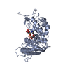
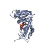
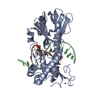
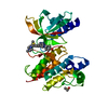
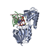
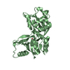
Mass: 32936.301 Da / Num. of mol.: 2
Fragment: Ligand Binding Domain (UNP residues 511-660, 776-915)
Source method: isolated from a genetically manipulated source
Details: THE PROTEIN CRYSTALLIZED IS THE EXTRACELLULAR LIGAND BINDING DOMAIN OF GluN3A. TRANSMEMBRANE REGIONS WERE GENETICALLY REMOVED AND REPLACED WITH A GLY-THR LINKE R THEREFORE, THE SEQUENCE ...Details: THE PROTEIN CRYSTALLIZED IS THE EXTRACELLULAR LIGAND BINDING DOMAIN OF GluN3A. TRANSMEMBRANE REGIONS WERE GENETICALLY REMOVED AND REPLACED WITH A GLY-THR LINKE R THEREFORE, THE SEQUENCE MATCHES DISCONTINUOUSLY WITH THE REFERENCE DATABASE (5 11-660 AND 776-915)
Source: (gene. exp.) Rattus norvegicus (Norway rat) / Gene: Grin3a / Plasmid: pET22 modified / Production host: Escherichia coli (E. coli) / Strain (production host): Origami B(DE3) / References: UniProt: Q9R1M7
#2: Chemical ChemComp-GOL / GLYCEROL / GLYCERIN / PROPANE-1,2,3-TRIOL


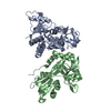
Mass: 92.094 Da / Num. of mol.: 2 / Source method: obtained synthetically / Formula: C3H8O3
#3: Water ChemComp-HOH / water


Mass: 18.015 Da / Num. of mol.: 579 / Source method: isolated from a natural source / Formula: H2O
Sequence detailsTHE PROTEIN CRYSTALLIZED IS THE EXTRACELLULAR LIGAND BINDING DOMAIN OF GlUN3A. TRANSMEMBRANE ...THE PROTEIN CRYSTALLIZED IS THE EXTRACELLULAR LIGAND BINDING DOMAIN OF GlUN3A. TRANSMEMBRANE REGIONS WERE GENETICALLY REMOVED AND REPLACED WITH A GLY-THR LINKER THEREFORE, THE SEQUENCE MATCHES DISCONTINUOUSLY WITH THE REFERENCE DATABASE (511-660 AND 776-915).

-
Experimental details

-
Experiment

ExperimentMethod: X-RAY DIFFRACTION / Number of used crystals: 1

-
Sample preparation

CrystalDensity Matthews: 2.37 Å3/Da / Density % sol: 48.06 %
Crystal growTemperature: 293 K / Method: vapor diffusion / pH: 5.4
Details: 10% PEG 6K, 0.1 M MES, 0.12 M NaCl, pH 5.4, VAPOR DIFFUSION, temperature 293K

-
Data collection

DiffractionMean temperature: 100 K
Diffraction sourceSource: SYNCHROTRON / Site: APS / Beamline: 22-ID / Wavelength: 1 Å
DetectorType: MARMOSAIC 300 mm CCD / Detector: CCD / Date: Feb 14, 2008 / Details: Double crystal
RadiationMonochromator: Si (111) / Protocol: SINGLE WAVELENGTH / Monochromatic (M) / Laue (L): M / Scattering type: x-ray
Radiation wavelengthWavelength: 1 Å / Relative weight: 1
ReflectionResolution: 1.68→30 Å / Num. all: 72079 / Num. obs: 72079 / % possible obs: 98.9 % / Observed criterion σ(F): 1 / Observed criterion σ(I): 1 / Redundancy: 7.1 % / Biso Wilson estimate: 18.2 Å2 / Rmerge(I) obs: 0.059 / Net I/σ(I): 27.9
Reflection shellResolution: 1.68→1.74 Å / Redundancy: 7.1 % / Rmerge(I) obs: 0.34 / Mean I/σ(I) obs: 6.9 / % possible all: 97.9

-
Processing

Software
NameVersionClassification
HKL-2000data collection
PHASERphasing
PHENIX(phenix.refine: 1.7.3_928)refinement
HKL-2000data reduction
HKL-2000data scaling
RefinementMethod to determine structure: MOLECULAR REPLACEMENT
Starting model: 2RC7
Resolution: 1.68→29.449 Å / SU ML: 0.18 / Cross valid method: THROUGHOUT / σ(F): 1.34 / Phase error: 16.43 / Stereochemistry target values: ML
RfactorNum. reflection% reflectionSelection details
Rfree0.184 3567 5.02 %RANDOM
Rwork0.1542 ---
obs0.1557 71098 98.9 %-
all-71098 --
Solvent computationShrinkage radii: 0.9 Å / VDW probe radii: 1.11 Å / Solvent model: FLAT BULK SOLVENT MODEL / Bsol: 40.474 Å2 / ksol: 0.4 e/Å3
Displacement parameters
Baniso -1Baniso -2Baniso -3
1--0.0764 Å20 Å2-0 Å2
2--0.44 Å20 Å2
3----0.3636 Å2
Refinement stepCycle: LAST / Resolution: 1.68→29.449 Å
ProteinNucleic acidLigandSolventTotal
Num. atoms4586 0 12 579 5177
Refine LS restraints
Refine-IDTypeDev idealNumber
X-RAY DIFFRACTIONf_bond_d0.014899
X-RAY DIFFRACTIONf_angle_d1.3076675
X-RAY DIFFRACTIONf_dihedral_angle_d12.2151828
X-RAY DIFFRACTIONf_chiral_restr0.08725
X-RAY DIFFRACTIONf_plane_restr0.007874
LS refinement shell
Resolution (Å)Rfactor RfreeNum. reflection RfreeRfactor RworkNum. reflection RworkRefine-ID% reflection obs (%)
1.68-1.7030.2641410.18522639X-RAY DIFFRACTION98
1.703-1.72740.24461410.17122605X-RAY DIFFRACTION98
1.7274-1.75310.19161390.16492650X-RAY DIFFRACTION98
1.7531-1.78050.22721530.16482644X-RAY DIFFRACTION98
1.7805-1.80970.22151340.15882637X-RAY DIFFRACTION98
1.8097-1.84090.16591360.15032658X-RAY DIFFRACTION98
1.8409-1.87440.18791380.14472677X-RAY DIFFRACTION98
1.8744-1.91040.18851370.15132659X-RAY DIFFRACTION98
1.9104-1.94940.1751390.14422647X-RAY DIFFRACTION98
1.9494-1.99180.20641460.14242647X-RAY DIFFRACTION99
1.9918-2.03810.18931550.142677X-RAY DIFFRACTION99
2.0381-2.08910.17151390.13982687X-RAY DIFFRACTION99
2.0891-2.14550.14821490.13692685X-RAY DIFFRACTION99
2.1455-2.20870.14531300.13432710X-RAY DIFFRACTION99
2.2087-2.27990.18531450.142668X-RAY DIFFRACTION99
2.2799-2.36140.18631450.14592721X-RAY DIFFRACTION99
2.3614-2.45590.19161370.1512722X-RAY DIFFRACTION99
2.4559-2.56760.19031420.15452687X-RAY DIFFRACTION99
2.5676-2.70290.2111340.15672746X-RAY DIFFRACTION100
2.7029-2.87210.18921480.15622718X-RAY DIFFRACTION100
2.8721-3.09360.17171460.15592756X-RAY DIFFRACTION100
3.0936-3.40450.17961530.16032761X-RAY DIFFRACTION100
3.4045-3.89620.16581470.1542769X-RAY DIFFRACTION100
3.8962-4.90510.16391420.1352818X-RAY DIFFRACTION100
4.9051-29.45370.20631510.22943X-RAY DIFFRACTION100
Refinement TLS params.

Method: refined / Refine-ID: X-RAY DIFFRACTION

IDL112)L122)L132)L222)L232)L332)S11 (Å °)S12 (Å °)S13 (Å °)S21 (Å °)S22 (Å °)S23 (Å °)S31 (Å °)S32 (Å °)S33 (Å °)T112)T122)T132)T222)T232)T332)Origin x (Å)Origin y (Å)Origin z (Å)
12.04170.24720.66961.33880.58292.1931-0.05250.11330.0757-0.1016-0.03990.2087-0.0476-0.1010.01010.0493-0.012-0.02040.0572-0.00020.0526-4.3774-2.8409-25.0859
21.8523-0.68360.01464.406-0.87452.6892-0.0586-0.33050.29560.5569-0.0231-0.2827-0.16080.34410.03480.0766-0.0167-0.00120.1624-0.03770.122716.99646.0122-1.214
31.9021-0.6403-0.56592.5059-0.13962.066-0.1435-0.2535-0.14550.19520.01610.04170.379-0.02780.07230.10280.00670.03770.09580.01520.07368.2764-3.61771.499
41.03130.02460.60460.3912-0.09551.7292-0.02570.13960.1101-0.05070.0474-0.0333-0.14770.1982-0.04920.0709-0.03160.01860.08120.0140.08459.93933.9424-17.004
51.47090.50180.26951.3410.31391.87690.035-0.28250.06790.0663-0.0221-0.1269-0.01650.3848-0.01280.070.01190.01340.189-0.00480.12144.7774-3.9175-7.1515
61.45980.2944-0.14861.15720.09461.4802-0.0893-0.2325-0.1979-0.00770.0263-0.14780.2620.1342-0.01050.10570.05410.0280.07940.03980.099737.7737-15.55-12.5293
71.6064-0.1573-0.20832.05610.66241.79810.02370.0861-0.0288-0.0892-0.0035-0.21590.04780.07150.00670.05930.021-0.00040.0572-0.00110.08944.6418-17.8414-36.5612
81.0220.09650.08631.5568-0.26261.4677-0.0977-0.0332-0.0463-0.02140.02370.06260.0518-0.09170.05750.06330.01790.01170.076-0.01260.09127.5657-11.1397-19.0342
Refinement TLS group
IDRefine-IDRefine TLS-IDSelection details
1X-RAY DIFFRACTION1(chain A and resid 4:143)
2X-RAY DIFFRACTION2(chain A and resid 144:164)
3X-RAY DIFFRACTION3(chain A and resid 165:198)
4X-RAY DIFFRACTION4(chain A and resid 199:292)
5X-RAY DIFFRACTION5(chain B and resid 4:64)
6X-RAY DIFFRACTION6(chain B and resid 65:142)
7X-RAY DIFFRACTION7(chain B and resid 143:249)
8X-RAY DIFFRACTION8(chain B and resid 250:294)

+
About Yorodumi

-
News

-
Feb 9, 2022. New format data for meta-information of EMDB entries

New format data for meta-information of EMDB entries

  • Version 3 of the EMDB header file is now the official format.
  • The previous official version 1.9 will be removed from the archive.

Related info.:EMDB header

External links:wwPDB to switch to version 3 of the EMDB data model

-
Aug 12, 2020. Covid-19 info

Covid-19 info

URL: https://pdbjlvh1.pdbj.org/emnavi/covid19.php

New page: Covid-19 featured information page in EM Navigator.

Related info.:Covid-19 info / Mar 5, 2020. Novel coronavirus structure data

+
Mar 5, 2020. Novel coronavirus structure data

Novel coronavirus structure data

Related info.:Yorodumi Speices / Aug 12, 2020. Covid-19 info

External links:COVID-19 featured content - PDBj / Molecule of the Month (242):Coronavirus Proteases

+
Jan 31, 2019. EMDB accession codes are about to change! (news from PDBe EMDB page)

EMDB accession codes are about to change! (news from PDBe EMDB page)

  • The allocation of 4 digits for EMDB accession codes will soon come to an end. Whilst these codes will remain in use, new EMDB accession codes will include an additional digit and will expand incrementally as the available range of codes is exhausted. The current 4-digit format prefixed with “EMD-” (i.e. EMD-XXXX) will advance to a 5-digit format (i.e. EMD-XXXXX), and so on. It is currently estimated that the 4-digit codes will be depleted around Spring 2019, at which point the 5-digit format will come into force.
  • The EM Navigator/Yorodumi systems omit the EMD- prefix.

Related info.:Q: What is EMD? / ID/Accession-code notation in Yorodumi/EM Navigator

External links:EMDB Accession Codes are Changing Soon! / Contact to PDBj

+
Jul 12, 2017. Major update of PDB

Major update of PDB

  • wwPDB released updated PDB data conforming to the new PDBx/mmCIF dictionary.
  • This is a major update changing the version number from 4 to 5, and with Remediation, in which all the entries are updated.
  • In this update, many items about electron microscopy experimental information are reorganized (e.g. em_software).
  • Now, EM Navigator and Yorodumi are based on the updated data.

External links:wwPDB Remediation / Enriched Model Files Conforming to OneDep Data Standards Now Available in the PDB FTP Archive

-
Yorodumi

Thousand views of thousand structures

  • Yorodumi is a browser for structure data from EMDB, PDB, SASBDB, etc.
  • This page is also the successor to EM Navigator detail page, and also detail information page/front-end page for Omokage search.
  • The word "yorodu" (or yorozu) is an old Japanese word meaning "ten thousand". "mi" (miru) is to see.

Related info.:EMDB / PDB / SASBDB / Comparison of 3 databanks / Yorodumi Search / Aug 31, 2016. New EM Navigator & Yorodumi / Yorodumi Papers / Jmol/JSmol / Function and homology information / Changes in new EM Navigator and Yorodumi

Read more