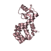
 Movie
Movie Controller

[English] 日本語
 Yorodumi
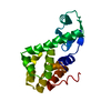
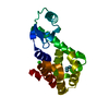
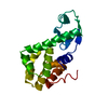
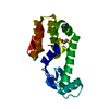
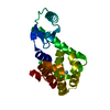
- PDB-149l: CONSERVATION OF SOLVENT-BINDING SITES IN 10 CRYSTAL FORMS OF T4 L... -
+ Open data

- Basic information
| Entry | Database: PDB / ID: 149l | ||||||
|---|---|---|---|---|---|---|---|
| Title | CONSERVATION OF SOLVENT-BINDING SITES IN 10 CRYSTAL FORMS OF T4 LYSOZYME | ||||||
 Components Components | T4 LYSOZYME | ||||||
 Keywords Keywords | HYDROLASE(O-GLYCOSYL) | ||||||
| Function / homology |  Function and homology information Function and homology informationviral release from host cell by cytolysis / peptidoglycan catabolic process / cell wall macromolecule catabolic process / lysozyme / lysozyme activity / host cell cytoplasm / defense response to bacterium Similarity search - Function | ||||||
| Biological species |  Enterobacteria phage T4 (virus) Enterobacteria phage T4 (virus) | ||||||
| Method |  X-RAY DIFFRACTION / Resolution: 2.6 Å X-RAY DIFFRACTION / Resolution: 2.6 Å | ||||||
 Authors Authors | Wilson, K. / Matthews, B.W. | ||||||
 Citation Citation | Journal: Protein Sci. / Year: 1994 Title: Conservation of solvent-binding sites in 10 crystal forms of T4 lysozyme. Authors: Zhang, X.J. / Matthews, B.W. #1: Journal: Nature / Year: 1988Title: Hydrophobic Stabilization in T4 Lysozyme Determined Directly by Multiple Substitutions of Ile 3 Authors: Matsumura, M. / Becktel, W.J. / Matthews, B.W. #2: Journal: J.Biol.Chem. / Year: 1989Title: Structural Studies of Mutants of T4 Lysozyme that Alter Hydrophobic Stabilization Authors: Matsumura, M. / Wozniak, J.A. / Sun, D. / Matthews, B.W. | ||||||
| History |
|
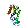
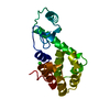
- Structure visualization
| Structure viewer | Molecule: MolmilJmol/JSmol |
|---|
- Downloads & links
-Download
| PDBx/mmCIF format | 149l.cif.gz | 43.9 KB | Display | PDBx/mmCIF format |
|---|---|---|---|---|
| PDB format | pdb149l.ent.gz | 31.3 KB | Display | PDB format |
| PDBx/mmJSON format | 149l.json.gz | Tree view | PDBx/mmJSON format | |
| Others |  Other downloads Other downloads |
-Validation report
| Arichive directory | https://data.pdbj.org/pub/pdb/validation_reports/49/149lftp://data.pdbj.org/pub/pdb/validation_reports/49/149l | HTTPS FTP |
|---|
-Related structure data


-Links
 PDBj
PDBj


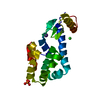
- Assembly
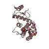
| Deposited unit | 
| ||||||||
|---|---|---|---|---|---|---|---|---|---|
| 1 |
| ||||||||
| Unit cell |
|
-Components
| #1: Protein | Mass: 18662.467 Da / Num. of mol.: 1 Source method: isolated from a genetically manipulated source Source: (gene. exp.) Enterobacteria phage T4 (virus) / Genus: T4-like viruses / Species: Enterobacteria phage T4 sensu lato / Plasmid: M13 / References: UniProt: P00720, lysozyme |
|---|---|
| #2: Water | ChemComp-HOH /  Mass: 18.015 Da / Num. of mol.: 38 / Source method: isolated from a natural source / Formula: H2O Mass: 18.015 Da / Num. of mol.: 38 / Source method: isolated from a natural source / Formula: H2O |
-Experimental details
-Experiment
| Experiment | Method: X-RAY DIFFRACTION |
|---|
- Sample preparation
| Crystal | Density Matthews: 2.56 Å3/Da / Density % sol: 51.89 % | ||||||||||||||||||||
|---|---|---|---|---|---|---|---|---|---|---|---|---|---|---|---|---|---|---|---|---|---|
| Crystal grow | *PLUS pH: 6.8 / Method: unknown | ||||||||||||||||||||
| Components of the solutions | *PLUS
|
-Data collection
| Radiation | Scattering type: x-ray |
|---|---|
| Radiation wavelength | Relative weight: 1 |
- Processing
| Software | Name: TNT / Classification: refinement | ||||||||||||||||||||||||||||||
|---|---|---|---|---|---|---|---|---|---|---|---|---|---|---|---|---|---|---|---|---|---|---|---|---|---|---|---|---|---|---|---|
| Refinement | Resolution: 2.6→10 Å / σ(F): 0 /
| ||||||||||||||||||||||||||||||
| Refinement step | Cycle: LAST / Resolution: 2.6→10 Å
| ||||||||||||||||||||||||||||||
| Refine LS restraints |
| ||||||||||||||||||||||||||||||
| Software | *PLUS Name: TNT / Classification: refinement | ||||||||||||||||||||||||||||||
| Refinement | *PLUS Highest resolution: 2.6 Å / Lowest resolution: 10 Å / Num. reflection all: 5904 / σ(F): 0 / Rfactor all: 0.192 | ||||||||||||||||||||||||||||||
| Solvent computation | *PLUS | ||||||||||||||||||||||||||||||
| Displacement parameters | *PLUS | ||||||||||||||||||||||||||||||
| Refine LS restraints | *PLUS
|
