 Movie
Movie Controller

[English] 日本語
 Yorodumi
- PDB-1jqu: Are Carboxy Terminii of Helices Coded by the Local Sequence or by... -
+ Open data

- Basic information
| Entry | Database: PDB / ID: 1jqu | ||||||
|---|---|---|---|---|---|---|---|
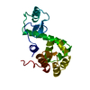
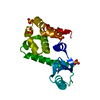
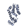
| Title | Are Carboxy Terminii of Helices Coded by the Local Sequence or by Tertiary Structure Contacts | ||||||
 Components Components | Lysozyme | ||||||
 Keywords Keywords | HYDROLASE / glycine helix terminii / Schellman motif / alpha-L motif | ||||||
| Function / homology |  Function and homology information Function and homology informationviral release from host cell by cytolysis / peptidoglycan catabolic process / cell wall macromolecule catabolic process / lysozyme / lysozyme activity / host cell cytoplasm / defense response to bacterium Similarity search - Function | ||||||
| Biological species |  Enterobacteria phage T4 (virus) Enterobacteria phage T4 (virus) | ||||||
| Method |  X-RAY DIFFRACTION / MOLECULAR REPLACEMENT / Resolution: 2.6 Å X-RAY DIFFRACTION / MOLECULAR REPLACEMENT / Resolution: 2.6 Å | ||||||
 Authors Authors | Sagermann, M. / Martensson, L.-G. / Baase, W.A. / Matthews, B.W. | ||||||
 Citation Citation | Journal: Protein Sci. / Year: 2002 Title: A test of proposed rules for helix capping: Implications for protein design Authors: Sagermann, M. / Martensson, L.-G. / Baase, W.A. / Matthews, B.W. #1: Journal: Science / Year: 1994 Title: Rules for alpha-helix termination by glycine. Authors: Aurora, R. / Srinivasan, R. / Rose, G.D. #2: Journal: Protein Folding / Year: 1980Title: The alpha-L Conformations at the Ends of Helices Authors: Schellman, C. #3: Journal: J.Mol.Biol. / Year: 1987Title: Structure of bacteriophage T4 lysozyme refined at 1.7 A resolution. Authors: Weaver, L.H. / Matthews, B.W. | ||||||
| History |
|
- Structure visualization
| Structure viewer | Molecule: MolmilJmol/JSmol |
|---|
- Downloads & links
-Download
| PDBx/mmCIF format | 1jqu.cif.gz | 140.2 KB | Display | PDBx/mmCIF format |
|---|---|---|---|---|
| PDB format | pdb1jqu.ent.gz | 110.8 KB | Display | PDB format |
| PDBx/mmJSON format | 1jqu.json.gz | Tree view | PDBx/mmJSON format | |
| Others |  Other downloads Other downloads |
-Validation report
| Arichive directory | https://data.pdbj.org/pub/pdb/validation_reports/jq/1jquftp://data.pdbj.org/pub/pdb/validation_reports/jq/1jqu | HTTPS FTP |
|---|
-Related structure data
| Related structure data | 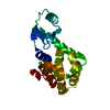 1llhC 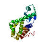 2lzmS C: citing same article ( S: Starting model for refinement |
|---|---|
| Similar structure data |


-Links
 PDBj
PDBj


- Assembly
| Deposited unit | 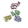
| ||||||||
|---|---|---|---|---|---|---|---|---|---|
| 1 | 
| ||||||||
| 2 | 
| ||||||||
| 3 | 
| ||||||||
| 4 | 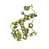
| ||||||||
| Unit cell |
|
-Components
| #1: Protein | Mass: 18555.312 Da / Num. of mol.: 4 / Mutation: C54T, C97A, W158L Source method: isolated from a genetically manipulated source Source: (gene. exp.) Enterobacteria phage T4 (virus) / Genus: T4-like viruses / Species: Enterobacteria phage T4 sensu lato / Gene: E / Plasmid: pHS1403 / Production host:  #2: Water | ChemComp-HOH / |  Mass: 18.015 Da / Num. of mol.: 29 / Source method: isolated from a natural source / Formula: H2O Mass: 18.015 Da / Num. of mol.: 29 / Source method: isolated from a natural source / Formula: H2O |
|---|
-Experimental details
-Experiment
| Experiment | Method: X-RAY DIFFRACTION / Number of used crystals: 1 |
|---|
- Sample preparation
| Crystal | Density Matthews: 2.67 Å3/Da / Density % sol: 53.93 % | ||||||||||||||||||||||||||||||||||||||||||
|---|---|---|---|---|---|---|---|---|---|---|---|---|---|---|---|---|---|---|---|---|---|---|---|---|---|---|---|---|---|---|---|---|---|---|---|---|---|---|---|---|---|---|---|
| Crystal grow | Temperature: 277 K / Method: vapor diffusion, hanging drop / pH: 6.75 Details: 30% PEG4000, PIPES buffer ph7.0, 0.2M LiSO4, pH 6.75, VAPOR DIFFUSION, HANGING DROP, temperature 277K | ||||||||||||||||||||||||||||||||||||||||||
| Crystal grow | *PLUS pH: 7 / Method: vapor diffusion | ||||||||||||||||||||||||||||||||||||||||||
| Components of the solutions | *PLUS
|
-Data collection
| Diffraction | Mean temperature: 298 K |
|---|---|
| Diffraction source | Source: ROTATING ANODE / Type: RIGAKU RU200 / Wavelength: 1.5418 Å |
| Detector | Type: XUONG-HAMLIN MULTIWIRE / Detector: AREA DETECTOR / Date: Nov 14, 1995 |
| Radiation | Monochromator: GRAPHITE / Protocol: SINGLE WAVELENGTH / Monochromatic (M) / Laue (L): M / Scattering type: x-ray |
| Radiation wavelength | Wavelength: 1.5418 Å / Relative weight: 1 |
| Reflection | Resolution: 2.6→34.9 Å / Num. all: 22095 / Num. obs: 22095 / % possible obs: 79 % / Observed criterion σ(F): 0 / Observed criterion σ(I): 0 / Redundancy: 3.42 % / Biso Wilson estimate: 36 Å2 / Rsym value: 0.057 / Net I/σ(I): 6.9 |
| Reflection shell | Resolution: 2.6→2.7 Å / Redundancy: 2.7 % / Mean I/σ(I) obs: 1.2 / Num. unique all: 3983 / Rsym value: 0.257 / % possible all: 71.5 |
| Reflection | *PLUS % possible obs: 78 % / Rmerge(I) obs: 0.057 |
- Processing
| Software |
| ||||||||||||||||||||
|---|---|---|---|---|---|---|---|---|---|---|---|---|---|---|---|---|---|---|---|---|---|
| Refinement | Method to determine structure: MOLECULAR REPLACEMENT Starting model: T4 Lysozyme 2LZM Resolution: 2.6→35 Å / Isotropic thermal model: Anisotropic / Cross valid method: THROUGHOUT / σ(F): 0 / Stereochemistry target values: Engh & Huber Details: Molecular replacement (direct rotation searches) was carried out with lysozyme search molecules with different hinge bending-angles. The starting model was subjected to several rounds of CNS ...Details: Molecular replacement (direct rotation searches) was carried out with lysozyme search molecules with different hinge bending-angles. The starting model was subjected to several rounds of CNS SA-refinement and subsequently refined with TNT. Positve density was observed around crystal contact sites. These features, presumably PEG molecules, could not be modeled unambigously and were therefore omitted from the final model.
| ||||||||||||||||||||
| Displacement parameters |
| ||||||||||||||||||||
| Refinement step | Cycle: LAST / Resolution: 2.6→35 Å
| ||||||||||||||||||||
| Refine LS restraints |
| ||||||||||||||||||||
| Refinement | *PLUS Highest resolution: 2.6 Å / σ(F): 0 / Rfactor all: 0.241 / Rfactor obs: 0.235 / Rfactor Rfree: 0.315 | ||||||||||||||||||||
| Solvent computation | *PLUS | ||||||||||||||||||||
| Displacement parameters | *PLUS | ||||||||||||||||||||
| Refine LS restraints | *PLUS
|
