 Movie
Movie Controller

[English] 日本語
 Yorodumi
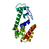
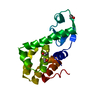
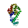
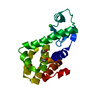
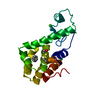
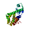
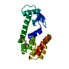
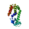
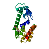
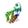
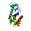
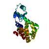
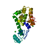
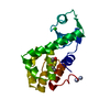
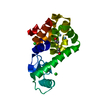
- PDB-1llh: ARE CARBOXY TERMINII OF HELICES CODED BY THE LOCAL SEQUENCE OR BY... -
+ Open data

- Basic information
| Entry | Database: PDB / ID: 1llh | |||||||||
|---|---|---|---|---|---|---|---|---|---|---|
| Title | ARE CARBOXY TERMINII OF HELICES CODED BY THE LOCAL SEQUENCE OR BY TERTIARY STRUCTURE CONTACTS | |||||||||
 Components Components | Lysozyme | |||||||||
 Keywords Keywords | HYDROLASE / helix terminii / schellman motif / alpha-l motif | |||||||||
| Function / homology |  Function and homology information Function and homology informationviral release from host cell by cytolysis / peptidoglycan catabolic process / cell wall macromolecule catabolic process / lysozyme / lysozyme activity / host cell cytoplasm / defense response to bacterium Similarity search - Function | |||||||||
| Biological species |  Enterobacteria phage T4 (virus) Enterobacteria phage T4 (virus) | |||||||||
| Method |  X-RAY DIFFRACTION / Isomorphous replacement / Resolution: 1.8 Å X-RAY DIFFRACTION / Isomorphous replacement / Resolution: 1.8 Å | |||||||||
 Authors Authors | Sagermann, M. / Martensson, L.-G. / Baase, W.A. / Matthews, B.W. | |||||||||
 Citation Citation | Journal: Protein Sci. / Year: 2002 Title: A test of proposed rules for helix capping: Implications for protein design Authors: Sagermann, M. / Martensson, L.-G. / Baase, W.A. / Matthews, B.W. #1: Journal: Science / Year: 1994 Title: Rules for alpha-helix termination by glycine. Authors: Aurora, R. / Srinivasan, R. / Rose, G.D. #2: Journal: Protein Folding / Year: 1980Title: The alpha-L Conformations at the Ends of Helices Authors: Schellman, C. #3: Journal: J.Mol.Biol. / Year: 1987Title: Structure of bacteriophage T4 lysozyme refined at 1.7 A resolution. Authors: Weaver, L.H. / Matthews, B.W. | |||||||||
| History |
|
- Structure visualization
| Structure viewer | Molecule: MolmilJmol/JSmol |
|---|
- Downloads & links
-Download
| PDBx/mmCIF format | 1llh.cif.gz | 46.8 KB | Display | PDBx/mmCIF format |
|---|---|---|---|---|
| PDB format | pdb1llh.ent.gz | 32.3 KB | Display | PDB format |
| PDBx/mmJSON format | 1llh.json.gz | Tree view | PDBx/mmJSON format | |
| Others |  Other downloads Other downloads |
-Validation report
| Arichive directory | https://data.pdbj.org/pub/pdb/validation_reports/ll/1llhftp://data.pdbj.org/pub/pdb/validation_reports/ll/1llh | HTTPS FTP |
|---|
-Related structure data
| Related structure data | 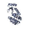 1jquC 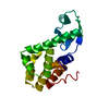 2lzmS C: citing same article ( S: Starting model for refinement |
|---|---|
| Similar structure data |


-Links
 PDBj
PDBj


- Assembly
| Deposited unit | 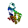
| ||||||||
|---|---|---|---|---|---|---|---|---|---|
| 1 |
| ||||||||
| Unit cell |
|
-Components
| #1: Protein | Mass: 18640.418 Da / Num. of mol.: 1 / Mutation: C54T, C97A, T157I Source method: isolated from a genetically manipulated source Source: (gene. exp.) Enterobacteria phage T4 (virus) / Genus: T4-like viruses / Species: Enterobacteria phage T4 sensu lato / Gene: E / Plasmid: pHS1403 / Production host:  | ||||
|---|---|---|---|---|---|
| #2: Chemical |   Mass: 35.453 Da / Num. of mol.: 2 / Source method: obtained synthetically / Formula: Cl Mass: 35.453 Da / Num. of mol.: 2 / Source method: obtained synthetically / Formula: Cl#3: Chemical | ChemComp-BME / |   Mass: 78.133 Da / Num. of mol.: 1 / Source method: obtained synthetically / Formula: C2H6OS Mass: 78.133 Da / Num. of mol.: 1 / Source method: obtained synthetically / Formula: C2H6OS#4: Water | ChemComp-HOH / |  Mass: 18.015 Da / Num. of mol.: 59 / Source method: isolated from a natural source / Formula: H2O Mass: 18.015 Da / Num. of mol.: 59 / Source method: isolated from a natural source / Formula: H2O |
-Experimental details
-Experiment
| Experiment | Method: X-RAY DIFFRACTION / Number of used crystals: 1 |
|---|
- Sample preparation
| Crystal | Density Matthews: 2.8 Å3/Da / Density % sol: 56.12 % |
|---|---|
| Crystal grow | Temperature: 277 K / Method: vapor diffusion, hanging drop / pH: 7.8 Details: 1.8M Phosphate, pH 7.8, VAPOR DIFFUSION, HANGING DROP, temperature 277K |
-Data collection
| Diffraction | Ambient pressure: 101 kPa / Mean temperature: 298 K |
|---|---|
| Diffraction source | Source: rotating-anode X-ray tube / Type: RIGAKU RU200 / Wavelength: 1.5418 Å / Target: Cu |
| Detector | Type: AREA DETECTOR / Detector: AREA DETECTOR / Date: Feb 15, 1995 / Details: Xuong-Hamlin |
| Radiation | Monochromator: graphite / Protocol: SINGLE WAVELENGTH / Monochromatic (M) / Laue (L): M / Scattering type: x-ray / Wavelength: 1.5418 Å |
| Radiation wavelength | Wavelength: 1.5418 Å / Relative weight: 1 |
| Reflection | Resolution: 1.8→26.44 Å / Num. all: 17775 / Num. obs: 17775 / % possible obs: 81 % / Observed criterion σ(F): 0 / Observed criterion σ(I): 0 / Redundancy: 2.37 % / Biso Wilson estimate: 16.36 Å2 / Rsym value: 0.04 / Net I/σ(I): 10.76 |
| Reflection shell | Resolution: 1.8→1.94 Å / Redundancy: 1.9 % / Rmerge(I) obs: 0.215 / Mean I/σ(I) obs: 1.9 / Num. unique all: 3101 / % possible all: 81 |
- Processing
| Software |
| |||||||||||||||||||||||||
|---|---|---|---|---|---|---|---|---|---|---|---|---|---|---|---|---|---|---|---|---|---|---|---|---|---|---|
| Refinement | Method to determine structure: Isomorphous replacement Starting model: PDB entry 2LZM Resolution: 1.8→27 Å / Isotropic thermal model: ANISOTROPIC / Cross valid method: THROUGHOUT / σ(F): 0 / σ(I): 0 / Stereochemistry target values: Engh & Huber Details: Residues 163 and 164 are missing in the electron density.
| |||||||||||||||||||||||||
| Displacement parameters |
| |||||||||||||||||||||||||
| Refinement step | Cycle: LAST / Resolution: 1.8→27 Å
| |||||||||||||||||||||||||
| Refine LS restraints |
|
