 Movie
Movie Controller


+ Open data

- Basic information
| Entry | Database: PDB / ID: 1epy | ||||||
|---|---|---|---|---|---|---|---|
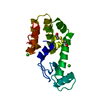
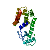
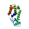
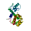
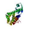
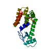
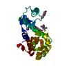
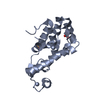
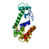
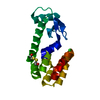
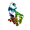
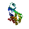
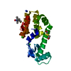
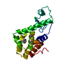
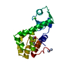
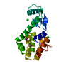
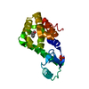
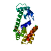
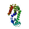
| Title | T4 LYSOZYME MUTANT, T21H/C54T/C97A/Q141H/T142H | ||||||
 Components Components | LYSOZYME | ||||||
 Keywords Keywords | HYDROLASE / metal binding | ||||||
| Function / homology |  Function and homology information Function and homology informationviral release from host cell by cytolysis / peptidoglycan catabolic process / cell wall macromolecule catabolic process / lysozyme / lysozyme activity / host cell cytoplasm / defense response to bacterium Similarity search - Function | ||||||
| Biological species |  Enterobacteria phage T4 (virus) Enterobacteria phage T4 (virus) | ||||||
| Method |  X-RAY DIFFRACTION / Resolution: 1.85 Å X-RAY DIFFRACTION / Resolution: 1.85 Å | ||||||
 Authors Authors | Wray, J.W. / Baase, W.A. / Ostheimer, G.J. / Zhang, X.-J. / Matthews, B.W. | ||||||
 Citation Citation | Journal: Protein Eng. / Year: 2000 Title: Use of a non-rigid region in T4 lysozyme to design an adaptable metal-binding site. Authors: Wray, J.W. / Baase, W.A. / Ostheimer, G.J. / Zhang, X.J. / Matthews, B.W. #1: Journal: Proc.Natl.Acad.Sci.USA / Year: 1989Title: Stabilization of Phage T4 Lysozyme by Engineered Disulfide Bonds Authors: Matsumura, M. / Becktel, W.J. / Levitt, M. / Matthews, B.W. | ||||||
| History |
|
- Structure visualization
| Structure viewer | Molecule: MolmilJmol/JSmol |
|---|
- Downloads & links
-Download
| PDBx/mmCIF format | 1epy.cif.gz | 47.3 KB | Display | PDBx/mmCIF format |
|---|---|---|---|---|
| PDB format | pdb1epy.ent.gz | 33.3 KB | Display | PDB format |
| PDBx/mmJSON format | 1epy.json.gz | Tree view | PDBx/mmJSON format | |
| Others |  Other downloads Other downloads |
-Validation report
| Arichive directory | https://data.pdbj.org/pub/pdb/validation_reports/ep/1epyftp://data.pdbj.org/pub/pdb/validation_reports/ep/1epy | HTTPS FTP |
|---|
-Related structure data


-Links
 PDBj
PDBj


- Assembly
| Deposited unit | 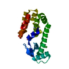
| ||||||||
|---|---|---|---|---|---|---|---|---|---|
| 1 |
| ||||||||
| Unit cell |
|
-Components
| #1: Protein | Mass: 18712.467 Da / Num. of mol.: 1 / Mutation: T21H,C54T,C97A,Q141H,T142H Source method: isolated from a genetically manipulated source Source: (gene. exp.) Enterobacteria phage T4 (virus) / Genus: T4-like viruses / Species: Enterobacteria phage T4 sensu lato / Production host:  |
|---|---|
| #2: Chemical | ChemComp-SO4 /   Mass: 96.063 Da / Num. of mol.: 1 / Source method: obtained synthetically / Formula: SO4 Mass: 96.063 Da / Num. of mol.: 1 / Source method: obtained synthetically / Formula: SO4 |
| #3: Chemical | ChemComp-CL /   Mass: 35.453 Da / Num. of mol.: 1 / Source method: obtained synthetically / Formula: Cl Mass: 35.453 Da / Num. of mol.: 1 / Source method: obtained synthetically / Formula: Cl |
| #4: Chemical | ChemComp-CO /   Mass: 58.933 Da / Num. of mol.: 1 / Source method: obtained synthetically / Formula: Co Mass: 58.933 Da / Num. of mol.: 1 / Source method: obtained synthetically / Formula: Co |
| #5: Water | ChemComp-HOH /  Mass: 18.015 Da / Num. of mol.: 98 / Source method: isolated from a natural source / Formula: H2O Mass: 18.015 Da / Num. of mol.: 98 / Source method: isolated from a natural source / Formula: H2O |
-Experimental details
-Experiment
| Experiment | Method: X-RAY DIFFRACTION / Number of used crystals: 1 |
|---|
- Sample preparation
| Crystal | Density Matthews: 2.92 Å3/Da / Density % sol: 57.86 % | ||||||||||||||||||||||||||||||||||||||||||||||||||||||
|---|---|---|---|---|---|---|---|---|---|---|---|---|---|---|---|---|---|---|---|---|---|---|---|---|---|---|---|---|---|---|---|---|---|---|---|---|---|---|---|---|---|---|---|---|---|---|---|---|---|---|---|---|---|---|---|
| Crystal grow | Temperature: 277 K / Method: vapor diffusion, hanging drop / pH: 7 Details: 12-16% PEG 8000, 10% isopropanol, sodium chloride, HEPES, 0.002 M cobalt chloride, pH 7.0, VAPOR DIFFUSION, HANGING DROP, temperature 4K | ||||||||||||||||||||||||||||||||||||||||||||||||||||||
| Crystal grow | *PLUS | ||||||||||||||||||||||||||||||||||||||||||||||||||||||
| Components of the solutions | *PLUS
|
-Data collection
| Diffraction | Mean temperature: 298 K |
|---|---|
| Diffraction source | Source: ROTATING ANODE / Type: RIGAKU RU200 / Wavelength: 1.5418 |
| Detector | Type: XUONG-HAMLIN MULTIWIRE / Detector: AREA DETECTOR / Date: Oct 11, 1997 |
| Radiation | Protocol: SINGLE WAVELENGTH / Monochromatic (M) / Laue (L): M / Scattering type: x-ray |
| Radiation wavelength | Wavelength: 1.5418 Å / Relative weight: 1 |
| Reflection | *PLUS Highest resolution: 1.8 Å / Num. obs: 19318 |
- Processing
| Software |
| ||||||||||||
|---|---|---|---|---|---|---|---|---|---|---|---|---|---|
| Refinement | Resolution: 1.85→20 Å / Stereochemistry target values: TNT
| ||||||||||||
| Refinement step | Cycle: LAST / Resolution: 1.85→20 Å
| ||||||||||||
| Refine LS restraints |
| ||||||||||||
| Software | *PLUS Name: TNT / Classification: refinement | ||||||||||||
| Refine LS restraints | *PLUS Type: t_plane_restr / Dev ideal: 0.019 |
