 Movie
Movie Controller


+ Open data

- Basic information
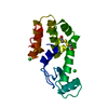
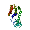
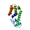
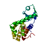
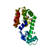
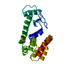
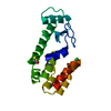
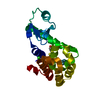
| Entry | Database: PDB / ID: 260l | ||||||
|---|---|---|---|---|---|---|---|
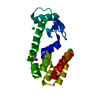
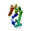
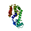
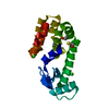
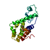
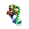
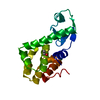
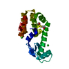
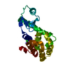
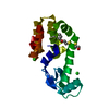
| Title | AN ADAPTABLE METAL-BINDING SITE ENGINEERED INTO T4 LYSOZYME | ||||||
 Components Components | PROTEIN (LYSOZYME) | ||||||
 Keywords Keywords | HYDROLASE / HYDROLASE (O-GLYCOSYL) / T4 LYSOZYME / METAL BINDING / PROTEIN ENGINEERING / PROTEIN DESIGN | ||||||
| Function / homology |  Function and homology information Function and homology informationviral release from host cell by cytolysis / peptidoglycan catabolic process / cell wall macromolecule catabolic process / lysozyme / lysozyme activity / host cell cytoplasm / defense response to bacterium Similarity search - Function | ||||||
| Biological species |  Enterobacteria phage T4 (virus) Enterobacteria phage T4 (virus) | ||||||
| Method |  X-RAY DIFFRACTION / MOLECULAR REPLACEMENT / Resolution: 1.8 Å X-RAY DIFFRACTION / MOLECULAR REPLACEMENT / Resolution: 1.8 Å | ||||||
 Authors Authors | Wray, J.W. / Baase, W.A. / Ostheimer, G.J. / Matthews, B.W. | ||||||
 Citation Citation | Journal: Protein Eng. / Year: 2000 Title: Use of a non-rigid region in T4 lysozyme to design an adaptable metal-binding site. Authors: Wray, J.W. / Baase, W.A. / Ostheimer, G.J. / Zhang, X.J. / Matthews, B.W. #1: Journal: Protein Sci. / Year: 1992Title: Structure of a Stabilizing Disulfide Bridge Mutant that Closes the Active Site Cleft of T4 Lysozyme Authors: Jacobson, R. / Matsumura, M. / Faber, H.R. / Matthews, B.W. #2: Journal: Science / Year: 1989Title: Control of Enzyme Activity by an Engineered Disulfide Bond Authors: Matsumura, M. / Matthews, B.W. #3: Journal: J.Mol.Biol. / Year: 1987Title: Structure of Bacteriophage T4 Lysozyme Refined at 1.7 Angstrom Resolution Authors: Weaver, L.H. / Matthews, B.W. | ||||||
| History |
|
- Structure visualization
| Structure viewer | Molecule: MolmilJmol/JSmol |
|---|
- Downloads & links
-Download
| PDBx/mmCIF format | 260l.cif.gz | 48.6 KB | Display | PDBx/mmCIF format |
|---|---|---|---|---|
| PDB format | pdb260l.ent.gz | 33.9 KB | Display | PDB format |
| PDBx/mmJSON format | 260l.json.gz | Tree view | PDBx/mmJSON format | |
| Others |  Other downloads Other downloads |
-Validation report
| Arichive directory | https://data.pdbj.org/pub/pdb/validation_reports/60/260lftp://data.pdbj.org/pub/pdb/validation_reports/60/260l | HTTPS FTP |
|---|
-Related structure data


-Links
 PDBj
PDBj


- Assembly
| Deposited unit | 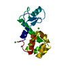
| ||||||||||
|---|---|---|---|---|---|---|---|---|---|---|---|
| 1 |
| ||||||||||
| Unit cell |
|
-Components
| #1: Protein | Mass: 18702.449 Da / Num. of mol.: 1 / Mutation: C54T,C97A,T21H,T142H Source method: isolated from a genetically manipulated source Details: T4 LYSOZYME MUTANT WITH CYS 54 REPLACED BY THR, CYS 97 REPLACED BY ALA, THR 21 REPLACED BY HIS, THR 142 REPLACED BY HIS Source: (gene. exp.) Enterobacteria phage T4 (virus) / Genus: T4-like viruses / Species: Enterobacteria phage T4 sensu latoDescription: BACTERIOPHAGE T4 (MUTANT GENE DERIVED FROM THE M13 PLASMID BY CLONING THE T4 LYSOZYME GENE) Cellular location: CYTOPLASM / Gene: GENE E FROM BACTERIOPHAGE T4 / Plasmid: PHS1403 / Gene (production host): T4 LYSOZYME / Production host:  | ||||||||||
|---|---|---|---|---|---|---|---|---|---|---|---|
| #2: Chemical |   Mass: 35.453 Da / Num. of mol.: 2 / Source method: obtained synthetically / Formula: Cl Mass: 35.453 Da / Num. of mol.: 2 / Source method: obtained synthetically / Formula: Cl#3: Chemical |   Mass: 58.693 Da / Num. of mol.: 2 / Source method: obtained synthetically / Formula: Ni Mass: 58.693 Da / Num. of mol.: 2 / Source method: obtained synthetically / Formula: Ni#4: Water | ChemComp-HOH / |  Mass: 18.015 Da / Num. of mol.: 107 / Source method: isolated from a natural source / Formula: H2O Mass: 18.015 Da / Num. of mol.: 107 / Source method: isolated from a natural source / Formula: H2OCompound details | STRUCTURE OF A T4 LYSOZYME MUTANT WITH AN ENGINEERED METAL BINDING SITE. THIS MUTANT DESIGNATED ...STRUCTURE OF A T4 LYSOZYME MUTANT WITH AN ENGINEERED | Nonpolymer details | NICKEL 2+ IS BOUND TO THE ENGINEERED PROTEIN LIGANDS HIS 21 AND HIS 142, WITH HOH 501-504 AS THE ...NICKEL 2+ IS BOUND TO THE ENGINEERED | Sequence details | RESIDUES 163 AND 164 ARE DISORDERED AND NOT OBSERVABLE IN ELECTRON DENSITY MAPS. THEREFORE THEY ARE ...RESIDUES 163 AND 164 ARE DISORDERED | |
-Experimental details
-Experiment
| Experiment | Method: X-RAY DIFFRACTION / Number of used crystals: 1 |
|---|
- Sample preparation
| Crystal | Density Matthews: 2.82 Å3/Da / Density % sol: 56.43 % / Description: STARTING MODEL WAS CYS-FREE WILDTYPE LYSOZYME | |||||||||||||||||||||||||||||||||||||||||||||||||||||||||||||||||||||||||||||
|---|---|---|---|---|---|---|---|---|---|---|---|---|---|---|---|---|---|---|---|---|---|---|---|---|---|---|---|---|---|---|---|---|---|---|---|---|---|---|---|---|---|---|---|---|---|---|---|---|---|---|---|---|---|---|---|---|---|---|---|---|---|---|---|---|---|---|---|---|---|---|---|---|---|---|---|---|---|---|
| Crystal grow | Temperature: 277 K / Method: vapor diffusion, hanging drop / pH: 7 Details: pH 7.0, VAPOR DIFFUSION, HANGING DROP, temperature 277K | |||||||||||||||||||||||||||||||||||||||||||||||||||||||||||||||||||||||||||||
| Components of the solutions |
| |||||||||||||||||||||||||||||||||||||||||||||||||||||||||||||||||||||||||||||
| Crystal grow | *PLUS Temperature: 4 ℃ | |||||||||||||||||||||||||||||||||||||||||||||||||||||||||||||||||||||||||||||
| Components of the solutions | *PLUS
|
-Data collection
| Diffraction | Mean temperature: 298 K |
|---|---|
| Diffraction source | Source: ROTATING ANODE / Type: RIGAKU RU200 / Wavelength: 1.5418 |
| Detector | Type: SDMS / Detector: AREA DETECTOR / Date: Jan 19, 1998 / Details: GRAPHITE MONOCHROMATOR |
| Radiation | Monochromator: NI FILTER/GRAPHITE / Protocol: SINGLE WAVELENGTH / Monochromatic (M) / Laue (L): M / Scattering type: x-ray |
| Radiation wavelength | Wavelength: 1.5418 Å / Relative weight: 1 |
| Reflection | Resolution: 1.8→20 Å / Num. obs: 19963 / % possible obs: 92 % / Redundancy: 2.5 % / Rmerge(I) obs: 0.031 |
| Reflection shell | Highest resolution: 1.8 Å / Rmerge(I) obs: 0.18 / Mean I/σ(I) obs: 2 |
- Processing
| Software |
| ||||||||||||||||||||||||||||||
|---|---|---|---|---|---|---|---|---|---|---|---|---|---|---|---|---|---|---|---|---|---|---|---|---|---|---|---|---|---|---|---|
| Refinement | Method to determine structure: MOLECULAR REPLACEMENT / Resolution: 1.8→20 Å / σ(F): 0 / Stereochemistry target values: TNT PROTGEO / Details: STARTING MODEL WAS CYS-FREE WILDTYPE
| ||||||||||||||||||||||||||||||
| Solvent computation | Solvent model: BABENET SCALING / Bsol: 790 Å2 / ksol: 1 e/Å3 | ||||||||||||||||||||||||||||||
| Refinement step | Cycle: LAST / Resolution: 1.8→20 Å
| ||||||||||||||||||||||||||||||
| Refine LS restraints |
| ||||||||||||||||||||||||||||||
| Software | *PLUS Name: TNT / Version: 5F / Classification: refinement | ||||||||||||||||||||||||||||||
| Refinement | *PLUS σ(F): 0 / Rfactor all: 0.19 | ||||||||||||||||||||||||||||||
| Solvent computation | *PLUS | ||||||||||||||||||||||||||||||
| Displacement parameters | *PLUS | ||||||||||||||||||||||||||||||
| Refine LS restraints | *PLUS
|
