 Movie
Movie Controller

[English] 日本語
 Yorodumi
- PDB-152l: CONSERVATION OF SOLVENT-BINDING SITES IN 10 CRYSTAL FORMS OF T4 L... -
+ Open data

- Basic information
| Entry | Database: PDB / ID: 152l | ||||||
|---|---|---|---|---|---|---|---|
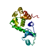
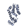
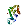
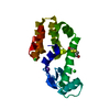
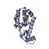
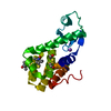
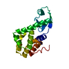
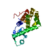
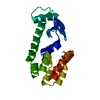
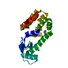
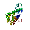
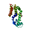
| Title | CONSERVATION OF SOLVENT-BINDING SITES IN 10 CRYSTAL FORMS OF T4 LYSOZYME | ||||||
 Components Components | T4 LYSOZYME | ||||||
 Keywords Keywords | HYDROLASE(O-GLYCOSYL) | ||||||
| Function / homology |  Function and homology information Function and homology informationviral release from host cell by cytolysis / peptidoglycan catabolic process / cell wall macromolecule catabolic process / lysozyme / lysozyme activity / host cell cytoplasm / defense response to bacterium Similarity search - Function | ||||||
| Biological species |  Enterobacteria phage T4 (virus) Enterobacteria phage T4 (virus) | ||||||
| Method |  X-RAY DIFFRACTION / Resolution: 2 Å X-RAY DIFFRACTION / Resolution: 2 Å | ||||||
 Authors Authors | Matsumura, M. / Weaver, L.H. / Matthews, B.W. | ||||||
 Citation Citation | Journal: Protein Sci. / Year: 1994 Title: Conservation of solvent-binding sites in 10 crystal forms of T4 lysozyme. Authors: Zhang, X.J. / Matthews, B.W. #1: Journal: To be PublishedTitle: A Covalent Enzyme-Substrate Intermediate with Saccharide Distorsion in a Mutant T4 Lysozyme Authors: Kuroki, R. / Weaver, L.H. / Matthews, B.W. #2: Journal: Protein Sci. / Year: 1992Title: Structure of a Stabilizing Disulfide Bridge Mutant that Closes the Active-Site Cleft of T4 Lysozyme Authors: Jacobson, R.H. / Matsumura, M. / Faber, H.R. / Matthews, B.W. #3: Journal: Biochemistry / Year: 1990Title: Structure of a Thermostable Disulfide-Bridge Mutant of Phage T4 Lysozyme Shows that an Engineered Cross-Link in a Flexible Region Does not Increase the Rigidity of the Folded Protein Authors: Pjura, P.E. / Matsumura, M. / Wozniak, J.A. / Matthews, B.W. #4: Journal: Nature / Year: 1989Title: Substantial Increase of Protein Stability by Multiple Disulphide Bonds Authors: Matsumura, M. / Signor, G. / Matthews, B.W. #5: Journal: J.Mol.Biol. / Year: 1987Title: Structure of Bacteriophage T4 Lysozyme Refined at 1.7 Angstroms Resolution Authors: Weaver, L.H. / Matthews, B.W. | ||||||
| History |
|
- Structure visualization
| Structure viewer | Molecule: MolmilJmol/JSmol |
|---|
- Downloads & links
-Download
| PDBx/mmCIF format | 152l.cif.gz | 46 KB | Display | PDBx/mmCIF format |
|---|---|---|---|---|
| PDB format | pdb152l.ent.gz | 32.2 KB | Display | PDB format |
| PDBx/mmJSON format | 152l.json.gz | Tree view | PDBx/mmJSON format | |
| Others |  Other downloads Other downloads |
-Validation report
| Arichive directory | https://data.pdbj.org/pub/pdb/validation_reports/52/152lftp://data.pdbj.org/pub/pdb/validation_reports/52/152l | HTTPS FTP |
|---|
-Related structure data

-Links
 PDBj
PDBj


- Assembly
| Deposited unit | 
| ||||||||
|---|---|---|---|---|---|---|---|---|---|
| 1 |
| ||||||||
| Unit cell |
|
-Components
| #1: Protein | Mass: 18634.461 Da / Num. of mol.: 1 Source method: isolated from a genetically manipulated source Source: (gene. exp.) Enterobacteria phage T4 (virus) / Genus: T4-like viruses / Species: Enterobacteria phage T4 sensu lato / Plasmid: M13 / References: UniProt: P00720, lysozyme |
|---|---|
| #2: Chemical | ChemComp-SO4 /   Mass: 96.063 Da / Num. of mol.: 1 / Source method: obtained synthetically / Formula: SO4 Mass: 96.063 Da / Num. of mol.: 1 / Source method: obtained synthetically / Formula: SO4 |
| #3: Water | ChemComp-HOH /  Mass: 18.015 Da / Num. of mol.: 52 / Source method: isolated from a natural source / Formula: H2O Mass: 18.015 Da / Num. of mol.: 52 / Source method: isolated from a natural source / Formula: H2O |
| Has protein modification | Y |
| Sequence details | SEQUENCE ADVISORY NOTICE DIFFERENCE BETWEEN SWISS-PROT AND PDB SEQUENCE. SWISS-PROT ENTRY NAME: ...SEQUENCE ADVISORY NOTICE DIFFERENCE |
-Experimental details
-Experiment
| Experiment | Method: X-RAY DIFFRACTION |
|---|
- Sample preparation
| Crystal | Density Matthews: 2.08 Å3/Da / Density % sol: 40.98 % | ||||||||||||||||||||
|---|---|---|---|---|---|---|---|---|---|---|---|---|---|---|---|---|---|---|---|---|---|
| Crystal grow | *PLUS pH: 8.5 / Method: unknown | ||||||||||||||||||||
| Components of the solutions | *PLUS
|
-Data collection
| Diffraction | Ambient pressure: 101 kPa / Mean temperature: 298 K |
|---|---|
| Diffraction source | Source: rotating-anode X-ray tube / Type: RIGAKU RU200 / Target: Cu |
| Detector | Type: AREA DETECTOR / Detector: AREA DETECTOR / Details: Xuong-Hamlin |
| Radiation | Monochromator: graphite / Protocol: SINGLE WAVELENGTH / Monochromatic (M) / Laue (L): M / Scattering type: x-ray / Wavelength: 1.5418 Å |
| Radiation wavelength | Relative weight: 1 |
- Processing
| Software |
| ||||||||||||||||||||||||||||||
|---|---|---|---|---|---|---|---|---|---|---|---|---|---|---|---|---|---|---|---|---|---|---|---|---|---|---|---|---|---|---|---|
| Refinement | Resolution: 2→50 Å / Rfactor obs: 0.168 / σ(F): 0 | ||||||||||||||||||||||||||||||
| Refinement step | Cycle: LAST / Resolution: 2→50 Å
| ||||||||||||||||||||||||||||||
| Refine LS restraints |
| ||||||||||||||||||||||||||||||
| Software | *PLUS Name: TNT / Classification: refinement | ||||||||||||||||||||||||||||||
| Refinement | *PLUS Highest resolution: 2 Å / Lowest resolution: 50 Å / σ(F): 0 / Rfactor all: 0.168 | ||||||||||||||||||||||||||||||
| Solvent computation | *PLUS | ||||||||||||||||||||||||||||||
| Displacement parameters | *PLUS | ||||||||||||||||||||||||||||||
| Refine LS restraints | *PLUS
|
