 Movie
Movie Controller

[English] 日本語
 Yorodumi
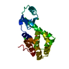
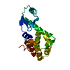
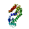
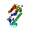
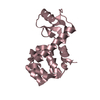
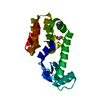
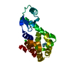
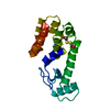
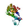
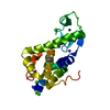
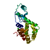
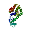
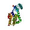
- PDB-1qth: THE INTRODUCTION OF STRAIN AND ITS EFFECTS ON THE STRUCTURE AND S... -
+ Open data

- Basic information
| Entry | Database: PDB / ID: 1qth | ||||||
|---|---|---|---|---|---|---|---|
| Title | THE INTRODUCTION OF STRAIN AND ITS EFFECTS ON THE STRUCTURE AND STABILITY OF T4 LYSOZYME | ||||||
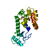
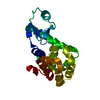
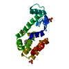
 Components Components | LYSOZYME | ||||||
 Keywords Keywords | HYDROLASE / STRAIN / STABILITY / MUTANT / T4 LYSOZYME | ||||||
| Function / homology |  Function and homology information Function and homology informationviral release from host cell by cytolysis / peptidoglycan catabolic process / cell wall macromolecule catabolic process / lysozyme / lysozyme activity / host cell cytoplasm / defense response to bacterium Similarity search - Function | ||||||
| Biological species |  Enterobacteria phage T4 (virus) Enterobacteria phage T4 (virus) | ||||||
| Method |  X-RAY DIFFRACTION / Resolution: 1.9 Å X-RAY DIFFRACTION / Resolution: 1.9 Å | ||||||
 Authors Authors | Liu, R. / Baase, W.A. / Matthews, B.W. | ||||||
 Citation Citation | Journal: J.Mol.Biol. / Year: 2000 Title: The introduction of strain and its effects on the structure and stability of T4 lysozyme. Authors: Liu, R. / Baase, W.A. / Matthews, B.W. | ||||||
| History |
|
- Structure visualization
| Structure viewer | Molecule: MolmilJmol/JSmol |
|---|
- Downloads & links
-Download
| PDBx/mmCIF format | 1qth.cif.gz | 77.3 KB | Display | PDBx/mmCIF format |
|---|---|---|---|---|
| PDB format | pdb1qth.ent.gz | 58.6 KB | Display | PDB format |
| PDBx/mmJSON format | 1qth.json.gz | Tree view | PDBx/mmJSON format | |
| Others |  Other downloads Other downloads |
-Validation report
| Arichive directory | https://data.pdbj.org/pub/pdb/validation_reports/qt/1qthftp://data.pdbj.org/pub/pdb/validation_reports/qt/1qth | HTTPS FTP |
|---|
-Related structure data
| Related structure data | 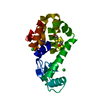 1qs5C 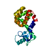 1qs9C 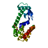 1qsbC 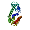 1qtbC 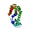 1qtcC  1qtdC C: citing same article ( |
|---|---|
| Similar structure data |


-Links
 PDBj
PDBj


- Assembly
| Deposited unit | 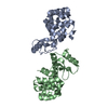
| ||||||||
|---|---|---|---|---|---|---|---|---|---|
| 1 | 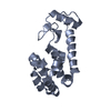
| ||||||||
| 2 | 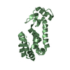
| ||||||||
| Unit cell |
| ||||||||
| Details | The biological assembly is a dimer constructed from chain A a symmetry partner generated by the noncrystallographic two-fold |
-Components
| #1: Protein | Mass: 18688.480 Da / Num. of mol.: 2 / Mutation: C54T, C97A, A98M Source method: isolated from a genetically manipulated source Source: (gene. exp.) Enterobacteria phage T4 (virus) / Genus: T4-like viruses / Species: Enterobacteria phage T4 sensu lato / Plasmid: M13 PHS1403 / Production host:  #2: Water | ChemComp-HOH / |  Mass: 18.015 Da / Num. of mol.: 113 / Source method: isolated from a natural source / Formula: H2O Mass: 18.015 Da / Num. of mol.: 113 / Source method: isolated from a natural source / Formula: H2O |
|---|
-Experimental details
-Experiment
| Experiment | Method: X-RAY DIFFRACTION / Number of used crystals: 1 |
|---|
- Sample preparation
| Crystal | Density Matthews: 2.26 Å3/Da / Density % sol: 45.51 % | |||||||||||||||||||||||||
|---|---|---|---|---|---|---|---|---|---|---|---|---|---|---|---|---|---|---|---|---|---|---|---|---|---|---|
| Crystal grow | Temperature: 277 K / Method: vapor diffusion, hanging drop / pH: 7.4 Details: PEG3.4K, magnesium chloride, Hepes, pH 7.4, VAPOR DIFFUSION, HANGING DROP, temperature 277K | |||||||||||||||||||||||||
| Crystal grow | *PLUS Method: unknown | |||||||||||||||||||||||||
| Components of the solutions | *PLUS
|
-Data collection
| Diffraction | Mean temperature: 298 K |
|---|---|
| Diffraction source | Source: ROTATING ANODE / Type: RIGAKU / Wavelength: 1.5418 |
| Detector | Type: RIGAKU RAXIS IIC / Detector: IMAGE PLATE / Date: Oct 29, 1997 |
| Radiation | Protocol: SINGLE WAVELENGTH / Monochromatic (M) / Laue (L): M / Scattering type: x-ray |
| Radiation wavelength | Wavelength: 1.5418 Å / Relative weight: 1 |
| Reflection | Resolution: 1.9→46.4 Å / Num. all: 25769 / Num. obs: 23171 / % possible obs: 89.92 % / Observed criterion σ(I): 0 / Biso Wilson estimate: 26.4 Å2 / Rmerge(I) obs: 0.048 |
| Reflection shell | Resolution: 1.9→2 Å / Rmerge(I) obs: 0.459 / Num. unique all: 3677 / % possible all: 86.4 |
| Reflection | *PLUS % possible obs: 85 % |
| Reflection shell | *PLUS % possible obs: 86.4 % |
- Processing
| Software |
| ||||||||||||||||||
|---|---|---|---|---|---|---|---|---|---|---|---|---|---|---|---|---|---|---|---|
| Refinement | Resolution: 1.9→20 Å / Stereochemistry target values: TNT PROTGEO
| ||||||||||||||||||
| Refinement step | Cycle: LAST / Resolution: 1.9→20 Å
| ||||||||||||||||||
| Refine LS restraints |
| ||||||||||||||||||
| Software | *PLUS Name: TNT / Classification: refinement | ||||||||||||||||||
| Refine LS restraints | *PLUS
|
